Cyclist’s Palsy

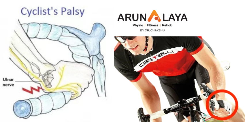
Cyclist’s palsy, also known by various names such as handlebar palsy, ulnar tunnel syndrome, ulnar nerve compression, Guyon Canal Syndrome (GCS), bicycler’s neuropathy, or tardy ulnar palsy, is an overuse injury that primarily affects the hands and fingers. It occurs when the nerves in the wrist or the side of the palm near the pinky finger become compressed due to repetitive stress.

Cause of cyclist palsy

  • Cyclist’s palsy can cause both motor and sensory symptoms.
  • The motor symptoms can include weak hand grip and difficulty using fingers for precise tasks.
  • Whereas the common sensory effects include numbness, tingling, and pain.

Although cyclist’s palsy is increasingly common, it is often underreported among cyclists. In fact, a study revealed that 7 out of 10 participants reported experiencing motor or sensory symptoms. Severe nerve injury can lead to paralysis or irreversible loss of sensation in the affected hand.

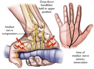
The pressure exerted on the handlebars during prolonged cycling can irritate the nerves in the palm. The highest pressure occurs where the median and ulnar nerves enter the hand, which corresponds to positions like “tops,” “ramps,” “hoods,” and “drops.” The “drops” position applies the most pressure on the ulnar nerve, while the “hoods” position applies slightly less pressure. The “tops” position places significant pressure on the palm at the base of the ring finger. The “drops” position can also cause excessive wrist extension, increasing pressure on the carpal tunnel. If a cyclist already has nerve compression at the neck or elbow, it can be more easily triggered at the palm, potentially leading to carpal tunnel syndrome or cubital tunnel syndrome.

Signs and Symptoms

  • include numbness, tingling, and sensory changes in the little finger and the ring finger on the side closest to the little finger
  • the palm in that area may also become numb, while there is no numbness on the back of the hand.
  • The symptoms can vary depending on the location of pressure. Sometimes manifesting as numbness or weakness, or a combination of both.
  • When the median nerve is affected, numbness and tingling occur on the palm side of the thumb, index, long, and ring fingers (on the side closest to the middle finger). But there is no numbness on the back of the hand.
  • Prolonged or severe pressure on the nerves can also weaken the associated muscles. Some cyclists may experience pain along with hand numbness.

Treatment

Limiting cycling is the most effective treatment for cyclist’s palsies. However, there are other measures that can allow cyclists to continue their activity while reducing the risk of exacerbating the condition. These include :

  • Limiting the length or distance of the ride
  • Having enough rest between longer cycling sessions
  • Changing positions of grip on the handlebars
  • Changing to a transverse handlebar
  • Adjusting the seat height
  • Using gloves to reduce or distribute pressure. The pressure can be reduced with foam or gel padding in the palm of the glove.

Top 3 Cyclist Palsy Exercises

In addition to these measures, exercises play a crucial role in long-term recovery and preventing recurrence of cyclist’s palsy. These exercises primarily focus on strengthening the muscles, ligaments, and tendons in the hands. The top three recommended exercises for cyclist’s palsy are as follows:

  1. Finger bending exercise: Begin by stretching your hand and then bend the fingers of the affected hand at a right angle, holding them in that position for approximately 10 seconds. Ensure that your fingers remain straight during the exercise. Repeat this process five times.
  2. Finger squeeze: Take a small object like a coin or a sheet of paper and squeeze it between two fingers, holding the grip for 10 seconds. Repeat this exercise five times for each set of fingers.
  3. Grip strengthening exercise: This exercise targets a weak hand grip. Squeeze a rubber ball with the affected hand and hold for 10 seconds and then release. Repeat 10 times, and that’s one set. Aim for 3 sets of 10 as you gradually build up grip strength.


Understanding Lateral Ligament Injury of the Ankle

Lateral ligament injury is among the most prevalent types of sports injuries addressed by physiotherapists. Men and women are estimated to suffer from lateral ankle sprains at roughly the same rates.

A lateral ligament injury of the ankle is a common injury that occurs when the ligaments on the outer side of the ankle are damaged. The most frequently affected ligaments are the anterior talofibular ligament (ATFL), the calcaneofibular ligament (CFL), and the posterior talofibular ligament (PTFL). These ligaments are responsible for providing stability to the ankle joint.

Lateral ankle sprains are also known as inversion or supination ankle sprains. The complex of ligaments on the lateral side of the ankle is torn by varied degrees as a result of a forced plantarflexion/inversion movement.

Individuals who sustain multiple recurrent ankle sprains have been documented to have functional and mechanical instability, as well as an increased risk of re-injury. Small fractures surrounding the ankle and foot (e.g. Pott’s fracture) and straining or rupture of the muscles around the ankle (e.g. calf, peroneii, tibialis anterior) should also be avoided even if they are the less prevalent causes of ankle pain.

Lateral Ankle Ligament Tear Types:

Lateral ligament injuries of the ankle can be classified into three main types based on the ligaments involved and the severity of the tear:

Anterior talofibular ligament (ATFL) injury: This is the most common type of lateral ligament injury and often occurs in isolation. It involves the stretching or tearing of the ATFL, which connects the talus bone to the fibula. It is commonly associated with ankle inversion injuries.

Calcaneofibular ligament (CFL) injury: The CFL is located slightly below the ATFL and connects the calcaneus (heel bone) to the fibula. CFL injuries typically occur along with ATFL injuries or in more severe sprains. In some cases, the ATFL and CFL can be injured together, resulting in a higher-grade ligament tear.

Posterior talofibular ligament (PTFL) injury: The PTFL is the least commonly injured ligament in lateral ligament injuries of the ankle. It connects the talus bone to the fibula at the back of the ankle. PTFL injuries usually occur in severe ankle sprains or high-energy trauma.

CAUSES

Ankle sprain: The most common cause of a lateral ligament injury is an ankle sprain, which typically occurs when the foot rolls inward, resulting in stretching or tearing of the ligaments on the outer side of the ankle.

Sports injuries: Activities that involve quick changes in direction, jumping, or running on uneven surfaces increase the risk of ankle sprains and ligament injuries.

Trauma: Direct trauma or impact to the ankle joint, such as a fall or a collision, can cause damage to the lateral ligaments.

CLINICAL FEATURES

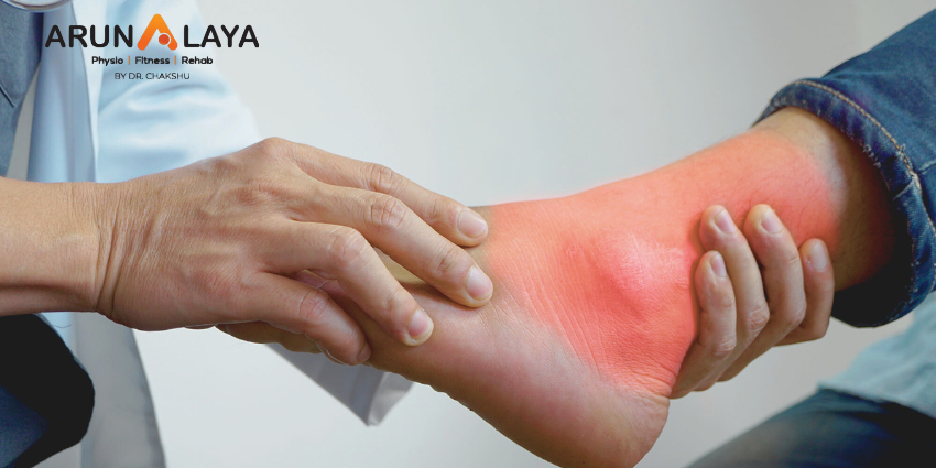
  • Pain on the outer side of the ankle
  • Swelling and bruising
  • Difficulty walking or bearing weight on the affected foot
  • Instability or a feeling of the ankle giving way
  • Limited range of motion in the ankle joint.

RISK FACTORS

Body mass index, slow eccentric inversion strength, quick concentric plantar flexion strength, passive inversion joint position sense, and peroneus brevis reaction time were all linked to an elevated risk of lateral ankle injury.

ASSESSMENT OF ANKLE JOINT

  • Amount of instability present by assessing the grade of the sprain;
  • Loss of Range of motion (ROM);
  • Loss of the muscle strength;
  • Level of reduced Proprioception.

OBSERVATION

Any symptoms of injury, inflammation, skin colour changes, or muscle atrophy or hypertrophy are noted. Following that, the foot and ankle are observed in two separate positions: non-weight bearing and weight bearing. Take note of the gait pattern, degree of limp (if any), and facial expression when bearing weight.

HISTORY

Mechanism of injury: A plantarflexion/inversion injury would indicate damage to the lateral ligament, whereas a dorsiflexion/eversion injury would indicate damage to the medial ligament. Previous history of injury on the same side will give clues as to whether the ankle was unstable to begin with, or that a previous injury wasn’t properly rehabilitated.

History of injury on the other side as well may indicate a biomechanical predisposition towards ankle injuries.

GRADES

Grade 1: Mild sprain with minimal stretching or tearing of the ligament fibers. There may be slight swelling, tenderness, and minimal loss of function.

Grade 2: Moderate sprain with partial tearing of the ligament fibers. This grade is characterized by increased swelling, bruising, pain, and difficulty walking or bearing weight on the affected ankle.

Grade 3: Severe sprain with complete tearing or rupture of the ligament. Significant swelling, bruising, pain, and instability are commonly observed. Walking or weight-bearing may be extremely difficult or impossible.

SPECIAL TEST

An anterior draw is performed to assess the ATFL and CFL integrity. The heel is grabbed with the ankle in plantarflexion, and the tibia is stabilised and dragged anteriorly.
The talar tilt is used to evaluate the integrity of the ATFL and CFL laterally, as well as the deltoid ligament medially. The heel is grabbed again, the tibia is stabilised, and the talus and calcaneus are pushed laterally and medially.
Beginning with a simple single leg stance, proprioception can be measured in a variety of more challenging methods. The patient can do it on the normal side first to give the therapist a sense of what is typical, and then try it on the injured side.

DIFFERENTIAL DIAGNOSIS

  • Ankle fracture (medial/lateral malleolus, distal tibia/fibular)
  • Damage to the medial ligament
  • Dislocated ankle
  • Other soft tissue damage (peroneal tendons, muscle strain)

TREATMENT

REDUCE PAIN AND SWELLING

The RICE regimen (Rest, Ice, Compression, and Elevation) can be used to minimise pain and swelling in the first 48-72 hours after an acute lateral ligament damage.

If weight bearing is too painful, the patient can use elbow crutches for 24 hours and remain non-weight bearing. However, it is critical to begin at least partial weight bearing as soon as possible, along with a regular heel-toe gait pattern, since this will help to reduce pain and swelling.

Gentle soft tissue massage and light stretches can be conducted to help with the clearance of oedema, as long as they are painless.

Range of motion exercises: These exercises aim to restore the normal range of motion in the ankle joint and may involve gentle ankle rotations, ankle pumps, and alphabet exercises.

Strengthening exercises: Strengthening the muscles around the ankle joint helps provide support and stability. Common exercises include calf raises, toe raises, ankle inversions and eversions (using resistance bands or manual resistance), and single-leg balance exercises.

Proprioceptive and balance exercises: These exercises improve the body’s awareness of joint position and enhance balance and stability. Examples include standing on one leg, balance board exercises, and wobble board exercises.

RETURN TO FUNCTIONAL ACTIVITY

  • Twisting
  • Jumping
  • Hopping on one leg
  • Running
  • Figure of 8 running

Before returning to full functional activity the patient should have full range of pain free movement in the ankle, normal strength and normal proprioception. If returning to sports, the athlete should be encouraged to wear an ankle brace or to tape the ankle for a further 6 months to provide external support.

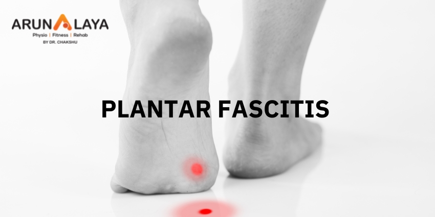
PLANTAR FASCITIS

Heel Pain medically known as Plantar fasciitis is a very common condition in today’s fast paced lifestyle. Apart from plantar fasciitis there are other causes of heel pain as well.

8 causes of Heel Pain

1.            Plantar fasciitis

2.            Heel spur

3.            Calf strain

4.            Achilles tendonitis

5.            Heel neuritis

6.            Heel bursitis

7.            Due to wearing high heels

8.            Stress fracture of the calcaneus

What are the symptoms of Plantar Fasciitis?

             Severe pain and swelling over heel

             Numbness or tingling in the heel

             You are unable to walk normally

             You can’t stand on the back of the foot

             Morning Pain in the heel

             Difficulty in walking

Diagnosis

The therapist will ask the patient how much walking and standing the patient does, what type of footwear is worn, and details of his/her medical history. Often this is enough to make a diagnosis.

Physical Examination

On physical examination, patients may walk with their affected foot in an equine position to avoid placing pressure on the painful heel. Palpation of the medial plantar calcaneal region will elicit a sharp, stabbing pain. Passive ankle/first toe dorsiflexion can cause discomfort in the proximal plantar fascia; it can also assess tightness of the Achilles tendon. Other causes of heel pain should be sought if history and physical examination findings are atypical for plantar fasciitis.

PHYSIOTHERAPY MANAGEMENT

Cryotherapy 15-20min. for reducing pain and swelling.

Ultrasonic therapy

TENS(Transcutaneous Electrical Nerve Stimulation)

Plantar fascia stretching

Calf stretches

Ankle strengthening exercises

Advanced Physiotherapy

Myofascial Release

Dry needling

Taping

IASTM

Prevention

Dont’s

Avoid wearing high heels.

Avoid prolonged standing.

Avoid running.

Avoid walking on uneven hard surfaces.

Do not walk bare foot even at home.

Avoid driving for long hours.

Do’s

Do stretch your calves muscle twice daily.

Do wear comfortable shoes with soft and spongy insoles. Use silica gel heel cushion.

Do take rest while prolonged standing or walking.

SHIN PAIN

Shin pain is common exercise related injury refers pain along the inner edge of shin bone (tibia) or sometimes on front of lower leg. Physical activity such as running, jumping or any vigorous sports activity can bring on shin pain, in case if you are starting at fresh or after a long time gap.

There can be some of the factors which can correlate as shin pain such as Stress fracture of tibia, Periostitis, Contusion injury, Compartment syndrome and MTSS (Medial Tibial Stress Syndrome). What is more common in athletes or exercise beginners is MTSS, known as shin splints where due to stress loading on tissues develop inflammation of muscles, tendons and bone tissue along the medial border of tibia (inner edge of shin bone).

Occurence

Generally, shin splints develop by overworking of muscles and periosteum (outer most layer of bone) by repetitive activities or bio-mechanical faults or environmental factors (exercise surface). Sudden changes in duration, intensity, and frequency of physical exercise can lead to shin splints.

Other factors which can contribute to shin splints are:-

             Flat feet or abnormal rigid arches can create abnormal stress on medial border of tibia (inner edge).

             Pain can be sharp, throbbing or dull.

             Pain occurs during and after exercise.

             Pain aggravates by touching on sore area or tender area.

If you ask, Should I take any medicine or go for radiological tests?

Oh No! Taking medicines such as anti-inflammatory and pain killers will not sort out your injury. Pills only kill the pain for some time, it’s momentarily relief and will not resolve your injury.

Secondly if you choose to do X-Ray, MRI etc., you will be wasting your time and money. Instead if you won’t find anything in radiological findings you can increase your anxiety level (beware of that).

Seek expert advice for the best treatment and go ahead as per their advice.

What will Sports physio do?

Assessment is a vital part for any injury or condition to be diagnosed. Sports physio will discuss about your pain, history and symptoms.

Shall do keen observation on your pain area

Shall palpate the tissues

Shall examine your movements

Do physical skill tests to evaluate and making correct diagnosis

Most importantly explaining to you the whole condition in the easiest ways that what it is.

How it occurred?

Why it happened?

What is the cure?

We the “Pain free zone” is a hub of specialized sports physio or expert in sports injuries. We will assess, diagnose and will treat you.

We have

-dynamic approach and multidimensional knowledge base which is patient centered

-clinical reasoning process that is embedded in problem solving approach

-central focus on movement assessment

-consistent virtues in caring and commitments towards patient.

“To have the effective treatment there is a need of correct assessment and diagnosis”

Coming to the topic again stress fracture, tendinitis, chronic exertional compartment syndrome can be detected by physical tests, movements and palpatory skills.

And we “PFZ” are highly expert in these.

Can I do something at home to relieve my pain before coming to you?

 Yeah why not?

             Do ice- use cold packs for 15-20 mins several times a day , but be careful about time duration

             Compression – wear elastic compressive bandage over pain area

             Proper shoes – wear good cushioning shoes to reduce stress on shin bone while walking.

             Avoid running on hard surface or either stop vigorous activity for some days.

If these can relieve my pain what all different you will do?

 You can have relief in pain by these but in future this pain can come again and can be more severe.

Why you wear sweaters and blazers in winters! Well you are feeling cold or last time you suffered from chills and fever.

Above mentioned are just home advices, treatment and solving your problem is way far.

 *If your pain is due to bio-mechanical fault such as over pronation, flat feet so ice and home remedies won’t work for you at all. That needs to be corrected

Pain free zone apply most advanced techniques in physiotherapy management

             Modalities

             Dry needling

             IASTM- Instrument Assisted Soft Tissue Mobilization

             Therapeutic Taping

             Manual Therapy

             Specific Exercise Program

             Return to Activities

Achilles Tendinitis

Achilles Tendinitis

In Greek mythology, a hero named Achilles, as a child, was dipped in the waters of the River Styx by his mother, by which means he became invulnerable, except for the part of his heel by which she held him (the proverbial “Achilles’ heel”) making it his only one weakness.

Read More

How does Physiotherapist help in Sports?

Sports Performance Enhancement
Sports Rehabilitation
With sports injuries having immediate and significant detrimental effects on your function, they are still a part of the game. Considering this, it is important to get you back in action. Our sports rehabilitation programme will help you recover and maximize your functional capacity, fitness and performance. Arunalaya Healthcare has a highly experienced team of physiotherapists who offer specialized treatment services for any sports injury.

Read More

Tingling and Numbness

Tingling and Numbness

Sometimes we can feel a sensation of pins and needles pricking us in any part of the body. In medical terms, this is known as Paresthesia. People generally notice these sensations in hands, feet, arms, and legs. If numbness and tingling persist and there’s no obvious cause for the sensations, it could be a symptom of a disease or injury, such as multiple sclerosis or carpal tunnel syndrome.

Read More