Understanding Lateral Ligament Injury of the Ankle

Lateral ligament injury is among the most prevalent types of sports injuries addressed by physiotherapists. Men and women are estimated to suffer from lateral ankle sprains at roughly the same rates.

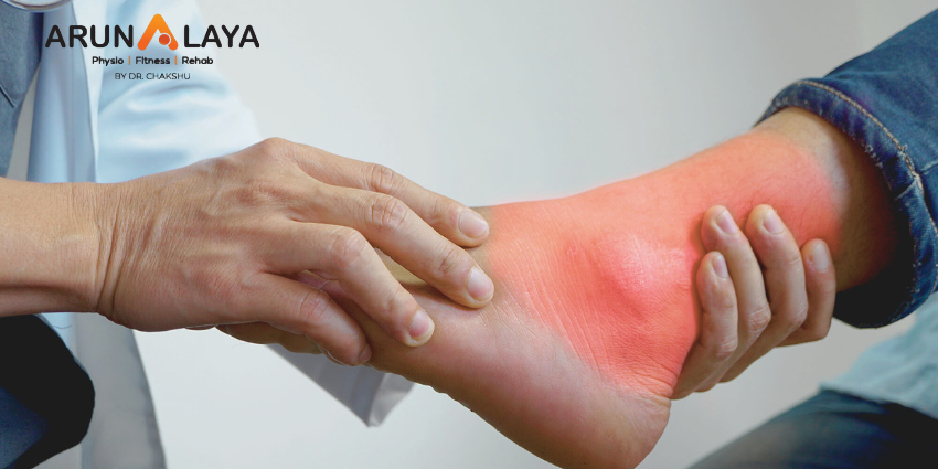
A lateral ligament injury of the ankle is a common injury that occurs when the ligaments on the outer side of the ankle are damaged. The most frequently affected ligaments are the anterior talofibular ligament (ATFL), the calcaneofibular ligament (CFL), and the posterior talofibular ligament (PTFL). These ligaments are responsible for providing stability to the ankle joint.

Lateral ankle sprains are also known as inversion or supination ankle sprains. The complex of ligaments on the lateral side of the ankle is torn by varied degrees as a result of a forced plantarflexion/inversion movement.

Individuals who sustain multiple recurrent ankle sprains have been documented to have functional and mechanical instability, as well as an increased risk of re-injury. Small fractures surrounding the ankle and foot (e.g. Pott’s fracture) and straining or rupture of the muscles around the ankle (e.g. calf, peroneii, tibialis anterior) should also be avoided even if they are the less prevalent causes of ankle pain.

Lateral Ankle Ligament Tear Types:

Lateral ligament injuries of the ankle can be classified into three main types based on the ligaments involved and the severity of the tear:

Anterior talofibular ligament (ATFL) injury: This is the most common type of lateral ligament injury and often occurs in isolation. It involves the stretching or tearing of the ATFL, which connects the talus bone to the fibula. It is commonly associated with ankle inversion injuries.

Calcaneofibular ligament (CFL) injury: The CFL is located slightly below the ATFL and connects the calcaneus (heel bone) to the fibula. CFL injuries typically occur along with ATFL injuries or in more severe sprains. In some cases, the ATFL and CFL can be injured together, resulting in a higher-grade ligament tear.

Posterior talofibular ligament (PTFL) injury: The PTFL is the least commonly injured ligament in lateral ligament injuries of the ankle. It connects the talus bone to the fibula at the back of the ankle. PTFL injuries usually occur in severe ankle sprains or high-energy trauma.

CAUSES

Ankle sprain: The most common cause of a lateral ligament injury is an ankle sprain, which typically occurs when the foot rolls inward, resulting in stretching or tearing of the ligaments on the outer side of the ankle.

Sports injuries: Activities that involve quick changes in direction, jumping, or running on uneven surfaces increase the risk of ankle sprains and ligament injuries.

Trauma: Direct trauma or impact to the ankle joint, such as a fall or a collision, can cause damage to the lateral ligaments.

CLINICAL FEATURES

  • Pain on the outer side of the ankle
  • Swelling and bruising
  • Difficulty walking or bearing weight on the affected foot
  • Instability or a feeling of the ankle giving way
  • Limited range of motion in the ankle joint.

RISK FACTORS

Body mass index, slow eccentric inversion strength, quick concentric plantar flexion strength, passive inversion joint position sense, and peroneus brevis reaction time were all linked to an elevated risk of lateral ankle injury.

ASSESSMENT OF ANKLE JOINT

  • Amount of instability present by assessing the grade of the sprain;
  • Loss of Range of motion (ROM);
  • Loss of the muscle strength;
  • Level of reduced Proprioception.

OBSERVATION

Any symptoms of injury, inflammation, skin colour changes, or muscle atrophy or hypertrophy are noted. Following that, the foot and ankle are observed in two separate positions: non-weight bearing and weight bearing. Take note of the gait pattern, degree of limp (if any), and facial expression when bearing weight.

HISTORY

Mechanism of injury: A plantarflexion/inversion injury would indicate damage to the lateral ligament, whereas a dorsiflexion/eversion injury would indicate damage to the medial ligament. Previous history of injury on the same side will give clues as to whether the ankle was unstable to begin with, or that a previous injury wasn’t properly rehabilitated.

History of injury on the other side as well may indicate a biomechanical predisposition towards ankle injuries.

GRADES

Grade 1: Mild sprain with minimal stretching or tearing of the ligament fibers. There may be slight swelling, tenderness, and minimal loss of function.

Grade 2: Moderate sprain with partial tearing of the ligament fibers. This grade is characterized by increased swelling, bruising, pain, and difficulty walking or bearing weight on the affected ankle.

Grade 3: Severe sprain with complete tearing or rupture of the ligament. Significant swelling, bruising, pain, and instability are commonly observed. Walking or weight-bearing may be extremely difficult or impossible.

SPECIAL TEST

An anterior draw is performed to assess the ATFL and CFL integrity. The heel is grabbed with the ankle in plantarflexion, and the tibia is stabilised and dragged anteriorly.
The talar tilt is used to evaluate the integrity of the ATFL and CFL laterally, as well as the deltoid ligament medially. The heel is grabbed again, the tibia is stabilised, and the talus and calcaneus are pushed laterally and medially.
Beginning with a simple single leg stance, proprioception can be measured in a variety of more challenging methods. The patient can do it on the normal side first to give the therapist a sense of what is typical, and then try it on the injured side.

DIFFERENTIAL DIAGNOSIS

  • Ankle fracture (medial/lateral malleolus, distal tibia/fibular)
  • Damage to the medial ligament
  • Dislocated ankle
  • Other soft tissue damage (peroneal tendons, muscle strain)

TREATMENT

REDUCE PAIN AND SWELLING

The RICE regimen (Rest, Ice, Compression, and Elevation) can be used to minimise pain and swelling in the first 48-72 hours after an acute lateral ligament damage.

If weight bearing is too painful, the patient can use elbow crutches for 24 hours and remain non-weight bearing. However, it is critical to begin at least partial weight bearing as soon as possible, along with a regular heel-toe gait pattern, since this will help to reduce pain and swelling.

Gentle soft tissue massage and light stretches can be conducted to help with the clearance of oedema, as long as they are painless.

Range of motion exercises: These exercises aim to restore the normal range of motion in the ankle joint and may involve gentle ankle rotations, ankle pumps, and alphabet exercises.

Strengthening exercises: Strengthening the muscles around the ankle joint helps provide support and stability. Common exercises include calf raises, toe raises, ankle inversions and eversions (using resistance bands or manual resistance), and single-leg balance exercises.

Proprioceptive and balance exercises: These exercises improve the body’s awareness of joint position and enhance balance and stability. Examples include standing on one leg, balance board exercises, and wobble board exercises.

RETURN TO FUNCTIONAL ACTIVITY

  • Twisting
  • Jumping
  • Hopping on one leg
  • Running
  • Figure of 8 running

Before returning to full functional activity the patient should have full range of pain free movement in the ankle, normal strength and normal proprioception. If returning to sports, the athlete should be encouraged to wear an ankle brace or to tape the ankle for a further 6 months to provide external support.
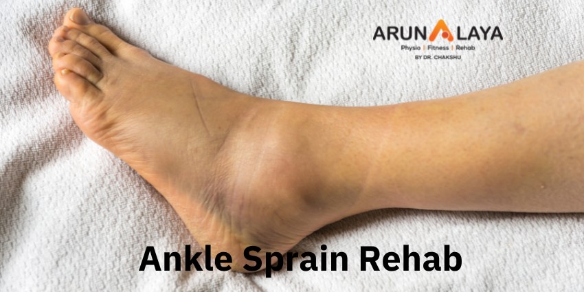
Ankle Sprain Rehab

Ankle Sprain Rehab

A sprained ankle is an injury that occurs when you roll, twist or turn your ankle in an awkward
way. This stretches or tear the ligaments which surrounds our ankle joint. Ankle sprain are
usually of 2 types . Medial and lateral ankle sprain . When your ankle gets twisted in innversion
then lateral sprain occur which is more common. When your ankle gets twisted in everson then
medial sprain occurs.
Ligaments which gets torned or stretched in lateral ankle sprain are
A) the Anterior talofibular ligament
B) Calcaneofibular ligament
C) Posterior talofibular ligament
Ligaments which gets affected in medial sprain are
A) Deltoid
Sign and symptoms of a sprained ankle( depends upon the severity of the sprain )
1) pain and tenderness
2) warmth
3) swelling
4) Restricted ROM
5) instability in the ankle
6) limp during walking
7) inability to bear weight on affected side
MANAGEMENT
grade 1 and grade 2 ankle sprains are treated conservatively. Surgical intervention is required
for grade 3.
The first protocol for sprained ankle is PRICE which means prevention from further injury, Rest,
Icing, Compression, Elevation .
1) patient can perform ankle ROM active movements within painfree limits to improve local
circulation
2) Cold compress or cryotherapy is unbeatable .
3) Ultrasonic on sprained ankle to reduce tenderness and inflammation
Strengthening exercises of ankle joint are done ones pain subcides.
POST OP rehab of a sprained ankle.
1) ankle toe pumps
2) Ankle ROM
3) Calf muscle pumping and stretching
4) isometric of of quads and hams
5) Toe raise
6) heel raise
7) toe curls
8) strengthening exercises with thearaband (resistance should be mild at the initiation )
9) walking to discard any gait abnormality
10) Strengthening exercises of the healthy limb
11) Balancing exercises
Teaching to patient to prevent from further injury and reoccurence is also necessary.

Tension headache

Tension headache

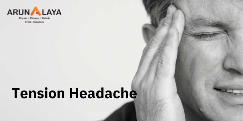
Headache caused by tensions feels like a mild to moderate pain on forhead , scalp and neck and
are often relived by taking painkillers and paracetamol.
CAUSES
Tension headaches usually occurs when cervical muscles becomes stiff and contracts in
response to stress, workload, any headinjury or anxiety. Adults are more prone to tension
headaches.
When our neck is in uncomfortable position for long duration it can cause headaches. Some
activities like typing work on laptops , mobiles or any activitiy which keeps our neck flexed may
trriger headaches.
Other triggers could be –
1) taking lot of caeffine
2) emotional stress
3) viral infections
4) alcohol or smoking habbits
5) ophthal problems
6) dental problems
7) dehydration
Several types of tension headaches are as follows
1) Sinus : In this type of headache pain is feel behind the eyebrow bone and cheekbones.
2) Cluster: throbbing pain is feel in and around one eye.
3) tension: pain feels like tight band over the forhead.
4) migraine: pain, nausea and visual changes are typical of classic form.
Treatment
Painkillers are the first line of treatment for tension headaches.
Common treatment include acetaminophen, NSAIDS
Physical therapy sessions including manual muscle release, dry needling electrical stimulation
modalities may provide relief from headaches.
Tension headache prevention
1) meditation
2) yoga and daily exercises
3) adequate fluid intake
4) correct posture at work
5) limit stress
6) eat balanced and healthy meals

HAGLUND DISEASE

Haglund Disease

It is a painful condition of the heel caused by mechanically induced inflammation of the
retrocalcaneal bursa, supracancaneal bursa and Achillies tendon.
The bump is usually due to an abnormality in foot function or bone position that creates shoe
friction around the natural bony prominence at the back of the heel.
The primary symptoms of Haglund deformity are
A) bump on the back of the heel
B) pain
C) swelling
D) callouses on and around the bump
Haglund deformity is believed to be caused by
A) high arches
B) tight achillies tendon
C) walking on outer edges of foot
D) Tight or poor fitting shoes
E) Abnormal biomechanics of foot due to joint misalignment.
Management of Hanglund deformity
First Non surgical treatment will be recommended .
Although there is no treatment to cure this deformity. Surgical interventions may also cause
bony bump again.
Some nonsurgical treatment choices include:
1) wear a shoe having rigid back.
2)placing heel lifts in shoes to help bring the heel up and avoid friction.
3)using heel pads inside the backs of shoes to help reduce irritation and friction on the heel.
4)people having high arches can use arch support inside the shoes.
5) analgesics can help in releiving pain symptom
6)cryotherapy can be done to reduce the inflammation and pain.
7) stretching exercises should be done for tight Achilles tendon.
8)avoiding climbing and running up hill. It will put more friction on bony prominence and will
alleviate pain.
9)use a soft cast that will reduce friction on the bone.
● If conservstive treatments will not relieve symptoms, the doctor may recommend surgery to
remove the part of the heel bone that sticks out.
Exercises therapy
According to the american college of foot and ankle surgeons,
1) heel raise
Stand with both feet flat on the floor, shoulder width apart. If you need support, steady yourself
with your hand on a wall or table. Hold for a few seconds, then lower the heels.Hold for 5–10
seconds, and then slowly lower the heels to the ground.
Repeat this exercise 10 times twice a day.
2) Heel drop
Begin by standing with one foot on a step and the heel raised up. Slowly lower the heel down
keeping the leg straight untill the foot is parallel to the ground but not further. Then come to the
starting position. Hold for 5 to 7 secs. Repeat to the normal position and repeat 10 times twice
a day.
3) heelcord stretch
While sitting on the ground, place the center of the towel around the ball of your foot while
holding each end of the towel with your hands. Lay on your back and raise your affected leg
while you pull on the towel ends untill you feel a stretch behind your leg.Hold for 7seconds.
Repeat this exercise 10 times twice a day.
4) towel stretch
Sit on the floor with both legs out .
Wrap a towel around one foot, holding both ends.Gently pull on the towel, pulling the ball of
the foot toward the body. There should be a gentle stretch in the calf muscle.Hold for 30
seconds and relax for 30 seconds. Repeat this exercise 10 times twice a day.
5)Towel scrunches
Keep a towel on the ground in front of the chair, then sit down on the chair with your heels on
the edge of thetowel. With one foot, reach out and use your toes to grab the towel, then pull the
towel towards you under your feet. Repeat this until you run out of towel, then repeat the whole
exercise 10 times thrice a day.

Joint cracking

Joint cracking

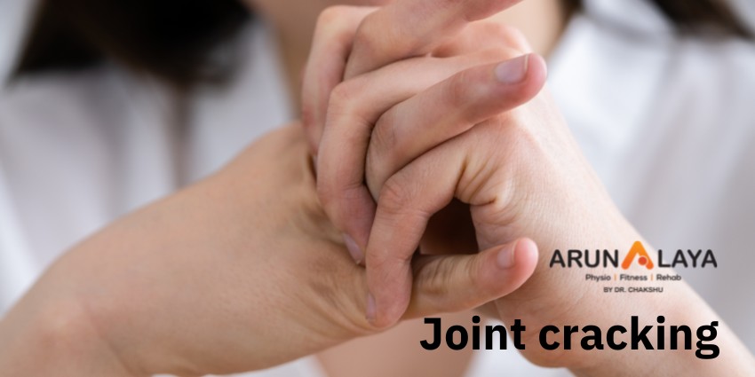
Cracking a joint is manipulating one’s joints to produce a distinct cracking or popping sound. It
is sometimes performed by physical therapists, chiropractors, osteopaths
The cracking mechanism and the resulting sound is caused by nitrogen cavitation bubbles
suddenly partially collapsing inside the joints. To be able to crack the same knuckle again
requires waiting about 15 minutes before the bubbles will be able to form again.
NOTE- “Cracking of joints doesnot believe to cause arthritis”.
CAUSES
In 2015, research showed that bubbles remained in the fluid after cracking, suggesting that
the cracking sound was produced when the bubble within the joint was formed, not when it
collapsed. In 2018, a team in France created a mathematical simulation of what happens in a
joint just before it cracks. The team concluded that the sound is caused by bubbles’ collapse,
and bubbles observed in the fluid are the result of a partial collapse. Due to the theoretical basis
and lack of physical experimentation, the scientific community is still not fully convinced of this
conclusion.
The snapping of tendons or scar tissue over a prominence (as in snapping hip syndrome) can
also generate a loud snapping or popping sound.
For many decades, the physical mechanism that causes the cracking sound as a result of
bending, twisting, or compressing joints was uncertain. Suggested causes included:
1)Cavitation within the joint—small cavities of partial vacuum form in the synovial fluid and then
rapidly collapse, producing a sharp sound.
2)Rapid stretching of ligaments.
3)Intra-articular (within-joint) adhesions being broken.
4)Formation of bubbles of joint air as the joint is expanded.
Why do people do it?
Studies show that as many as 54 percent of people crack their knuckles. They do it for a lot of
reasons, including:
Sound. Some people like hearing the sound knuckle cracking makes.
The way it feels. Some people think cracking their knuckles makes more room in the joint, which
relieves tension and increases mobility. However, although it may feel like there’s more room,
there’s no evidence that there actually is.
Nervousness. Just like wringing your hands or twirling your hair, cracking your knuckles may be a
way to occupy your hands when you’re nervous.
Stress. Some people who are stressed need to take it out on something. Cracking knuckles may
allow for diversion and release without actually causing harm.
Habit. Once you start cracking your knuckles for any of these reasons, it’s easy to keep doing it
until it happens without even thinking about it. When you find yourself unconsciously cracking
your knuckles many times a day, it’s become a habit. People who do it five times a day or more
are called habitual knuckle crackers.
Tips to stop cracking
1)Although cracking your knuckles isn’t harming you, it may be distracting to people around you.
You might find it difficult to stop if it’s become a habit.
2) Some tips that might help you break the habit:
3)Think about why you crack your knuckles and address any underlying issues.
4)Find another way to relieve stress, such as deep breathing, exercise, or meditation.
5)Occupy your hands with other stress relievers, such as squeezing a stress ball or rubbing a
worry stone.
6)Become aware of each time you crack your knuckles and consciously stop yourself

SHORTNESS OF BREATH

Shortness of Breath

Introduction- shortness of breath is the uncomfortable sensation of not getting enough air
to breathe. This may occur while walking, climbing stairs, running or even when sitting still. It
is also termed as dyspnoea. Sometimes it can be harmless as the result of exercise or nasal
congestion. In other situation, it may be a sign of a more serious heart or lung diseases.
CAUSES –
1) Anaphylaxis ( allergic reaction)
2) Asthma
3) Carbonmonooxide poisoning
4) COPD
5)Coronavirus disease 2019
6) Heart attack
7)Heart arrhythmia
8) Pneumonia
9) Pneumothorax
10) Pulmonary embolism
Diagnosis
1) Blood tests- Arterial blood gases and blood oxygen saturation may be measured.
2) Exercises test- blood pressure, heart rate and changes in breathing rate can be measured
during exercises and rest
3) Electrocardiogram- An ECG records the electrical activity of the heart and shows abnormal
rhthym.
4) Echocardiogram- An echo uses ultrasound waves to produce a moving pictures of the heart
and heart valves
5) Chest x rays- An x ray will help assess lung conditions
6)Ct scan of the chest.
Severity of dyspnea
Modified Medical Research council dyspnoea scale.
Grade 0- strenous exercises
Grade 1- hurrying up or walking up a hill
Grade 2- walks slower than people of same age or stops for taking breath
Grade 3- stops for breath after walking 100m
Grade 4- breathless while dressing and underdressing breathless to leave the home.
Types of dyspnea
Orthopnea- it is the feeling of dyspnea in the recumbent position, releived by sitting or standing.
Paroxysmal nocturnal dyspnea- its a sensation of dyspnea that awakens the patient, often after
1 or 2 hours of sleep, and is usually relieved in the upright position.
Trepopnea – it is a sensation of dyspnoea that occurs in one lateral decubitus position as
opposed to the other.
Platypnea- it is a sensation of dyspnoea that occurs in the upright position and is releived with
recumbency.
Management
Treatment will depend upon the cause of the problem.
In severe cases , supplemental oxygen will be needed . Those with asthma or copd may have an
inhaled rescue bronchodilator to use when necessary.
If dyspnoea is linked to asthma, it typically responds well to medications such as bacterial
pneumoniae , antibiotics can bring relief.
Other medication such as opiates, NSAIDS and anti- anxiety drugs can also be effective.
Physiotherapy management
Physiotherapist can offer a variety of treatments to both adults and children who suffer from
shortness of breath, giving them back the independence and control of their symptoms. Some
of the treatments and techniques used are:
1) Breathing techniques
2) Manual techniques
3) Postural drainage
4) lifestyle advice
5) flutter valve
6) incentive spirometery
7) ACBT
Breathing exercises1) pursed lip breathing helps empty the lungs of dead spaces air that occurs in COPD.
To perform pursed lip breathing:
1) relax your neck and shoulder muscles.
2) slowly breathe in through your nose for two counts, keeping your mouth closed.
3) purse your lips as if you are about to whistle.
4) breathe out slowly and gently through your pursed lips to the count of four.
Sitting forward supported by a table.
1) Sit in a chair with your feet flat on the floor, facing a table.
2) lean your chest slightly forward and rest your arms on the table .
3) Rest your head on your forearms or on a pillow.
Diaphragmatic breathing
1) Sit in a chair with bent knees and relaxed shoulders, head and neck.
2) place your hand on your belly.
3) Breathe in slowly through your nose . You should feel your belly moving under your hand.
4) As you exhale, tighten your muscles . You should feel your belly fall inward. Breathe out
through your mouth with pursed lips.
5) Put more emphasis on the exhale than the inhale. Keep exhaling for longer than usual before
slowly inhaling again.
6) Repeat for about mins.

What is “Flat Foot” Actually ?

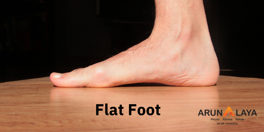
Flat foot is a foot deformity in which there is loss of medial longitudinal arch. It is also termed as
pes planus. Pes planus may be lifelong, or acquired through time. In this condition the entire sole
of the foot is in contact or near contact with the ground while standing.
flat foot is normal in infants and toddlers, because the foot’s arch hasn’t yet developed. Most
people’s arches develop throughout childhood, but some people never develop arches. This is a
normal variation in foot type, and people without arches may or may not have problems.
Some children have flexible flatfoot, in which the arch is visible when the child is sitting or
standing on tiptoes, but disappears when the child stands. Most children outgrow flexible
flatfoot without problems. Arches can also fall over time. Years of wear and tear can weaken the
tendon that runs along the inside of your ankle and helps support your arch.
Causes
Infants & children
Flat feet are common in children and are often caused by:
1)Heredity
2)Laxity of ligaments
3)Tight Achilles tendon
4)Lack of foot exercise
Typically a child’s arches begin developing in infancy and progress to normal arches in line with
normal growth patterns.
Adults can develop flat feet through injury, tight Achilles tendon, abnormal joint formation,
continued stresses on the foot and its arch, or simply as they age.
Some of the most common causes of flat feet in adults are:
1)Achilles Equinus contracture
2)Coalition of rearfoot joints
3)Failed or injured tendons
4)Arthritis
5)Marfan syndrome
6)Diabetes
7)Obesity
8)Pregnancy
9)Overuse & strain
10)Injury & fractures
The most identifiable symptoms and characteristics of flat feet are the decrease or lack of
arches in your feet (especially when weight bearing) and pain / fatigue along the inner side of
your feet and arches.Some issues caused by flat feet include:
1)Inflammation of soft tissue
2)Foot, arch, and leg fatigue
3)Heel, foot, and ankle pain
4)Knee, hip, and lower back pain
5)Rolled-in ankles
6)Abnormal walking patterns
7)Shin splints
8)Bunions
9)Hammertoe
10)Arthritis
11)Plantar fasciitis

Management of flat foot treatment
Treatment of flat feet will be started when you will start feeling pain or other foot, ankle
abnormality .
For children
At Advanced Foot & Ankle our podiatric specialists will conduct both a clinical exam and an
X-ray study to determine the type and cause of most flatfoot deformity.
Most cases of flat feet in children are genetic. However their feet are flexible in nature and can
be treated with:
1)Custom made insoles
2)Braces
3)Appropriate footwear
4)Exercise
5)Surgery – often the best treatment for preventing major developmental complications in the
future.
For Adults
Non-surgical treatment options for deterring the development of flat feet and its symptoms are:
1)Custom orthotics
2)Bracing & supports
3)Supportive footwear
4)Exercises
5)Physical Therapy
6)Weight loss
7)Daily activity modifications
8)Medication
9)Rest
Physiotherapy management will help to regain mobility of the foot, Strengthen the surrounding
muscles .Regain foot and ankle control Provide initial relief of pain by using ice pack wrapped
in a damp towel and placed on the foot or area of pain for 10-15 minute can help to reduce
soreness. Some patients prefer moist heat for pain relief.
1)Modalities : Heat and cold therapy is applied to enhance relaxation and reduces pain. Ultra
sound and pulsed electrical stimulation can also be used to relieve the pain. Electric stimulation
will helps to improve the blood circulation, thus enhancing the healing process and reducing any
swelling or discomfort.
2) Exercises: toe clawing exercises, arch muscle strengthening exercises, heel cord stretching,
calf and hammstring stretching, toe spreading exercises, toe walking, ankle ROM, Dorsiflexion
and plantar flexion of foot, gripping exercises. Releasing manually any tight muscles will also
develop arches and will bring the foot in shape. Kinesio taping to support and to avoid any
unwanted movement of muscle.
Advance techniques like dry needling on tendon and on trigger points will ease the pain .
If conservative treatment won’t help much in treating flat foot sign and symptoms then another
choice of treatment will be surgical.
Surgical interventions includes
1) tendon transfer
2) osteotomies
3) Arthrodesis.

How Physio can Help with Asthma?

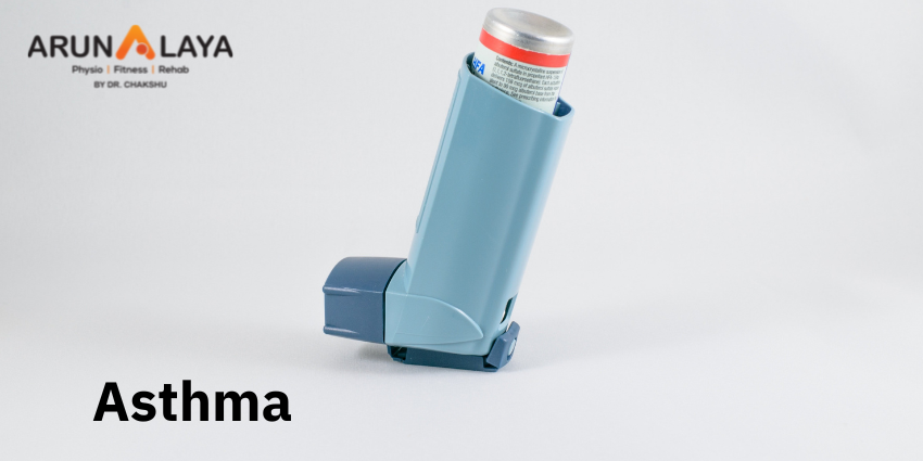
Asthma is a long term condition affecting children and adults. The air passage in the lungs
becomes narrow due to inflammation and tightening of the muscles around the small airways.
This causes asthma symptoms: cough, wheeze, shortness of breath and chest tightness. These
symptoms are intermittent and are often worse at night or during exercise. Other common
“triggers” can make asthma symptoms worse. Triggers vary from person to person, but can
include viral infections (colds), dust, smoke, fumes, changes in the weather, grass and tree
pollen, animal fur and feathers, strong soaps, and perfume.
Symptoms of asthma
1) Coughing at night, laughing or during exercise.
2) tightness in the chest
3) shortness of breath
4) difficulty talking
5) anxiety and panicking
6) fatigue, chest pain
7) rapid breathing
8) sleep interruptions
9)confusion
10)dizziness, pale lips and fingernails
Triggers include:
Respiratory infections, exercise, allergens, pests, enviornmental irritants, intense emotions,
extreme weather conditions.
Diagnosing asthma:
Pulmonary function test confirms the asthma. This can detect the stenosis in the lumen of
airways. Asthma is usually suspected by a healthcare provider based on a pattern of symptoms
and response to medicine called a bronchodilator that can releif the squeezing of the muscles
around the airways. Blood test to check for increased levels of eosinophils and immunoglobulin
E .
Management of asthma:
1) Bronchodilators that relax muscles around the airways.
2) Antibiotics to fight for any bacterial pneumonia and bronchitis.
3) Anti- inflammatory medications such as inhaled corticosteroids for long term, and oral
steroids for acute attacks
PHYSIOTHERAPY MANAGEMENT
Main aim of physiotherapy management will be to ease breathing and remove lung secretions
through chest physiotherapy.
Instructions are given from the respiratory therapist to how to cope up with the dyspnoea.
1) Decreasing breaths taken ( reducing respiratory flow).
2) taking smaller breaths ( reducing tidal volume)
3) Deep breathing ( diaphragmatic breathing through use of abdominal muscles and lower
throacic chest movements).
4) pursed lip breathing . Deep inhale from nose and exhaling through mouth like blowing air out.
5) relaxation plays vital role in managing asthma attacks. Sitting in semi flexed position keeping
4 to 5 pillows on the back for support will ease breathing .
6) deep breathing techniques. Inhale deep for 4 sec hold for 5 sec and blow out through mouth
slowly in 6 sec.
7) removal of secretions is very important as it triggers cough. Removal of chest secretions
through
a) percussions ( frequency should be more than intensity, intensity should be extreme low in
osteoporotic patients, or use vibrators in such patients)
b) shaking
c) vibrations
d) postural drainage
e) FET by huff cough.
8) Range of motion exercises for bed ridden patients to avoid any contractures.
9) Educate the patient about use of bronchodilators and breathing exercises.
10) correct posture in standing and sitting which will assist in the management of asthma
attacks by allowing the chest to expand appropriately and lungs to function optimally.
INSPIRATORY MUSCLES TRAINING: it can be trained for both strength and endurance
with an external resistive device. Exercise induced bronchoconstriction as well as chronic
bronchoconstriction in asthmatics is associated with increased inspiratory muscle work. It is
reasonable to suggest that increasing the strength of the inspiratory muscles in people with
asthma may reduce the intensity of dyspnea and improves exercise tolerance.
Breathing exercises, inspiratory muscle training, physical training and airway clearence are the
most relevant treatment options for asthmatic patients.

WHATSAPP THUMB SYNDROME

Whatsapp..hottest craze and necessity in today’s scenario. If we talk about youngsters their friends and group chatting and if we talk work then discussions on projects makes you involve in typing on mobile through your thumb resulting in pain in and around thumb.

The causative factor for Whatsapp syndrome is repetitive use of thumb musculature leading overuse strain. The muscles of thumb like flexor pollicis, Opponens pollicis and abductor pollicis gets repeated action leading to formation of trigger points.

Consequences –

Overuse syndrome can give rise to degeneration in the bones and joints.

Osteoarthritis in chronic cases can be caused.

How to avoid –

Pain in the thumb can be avoided by intermittent rest period.

Stretching the thumb musculature.

How Physiotherapy can help –

Muscle strain caused by repeated action of typing can be resolved by many advanced therapeutic techniques in physiotherapy like Dry needling, myofascial release and laser therapy.

Stretching of the musculature and taping the structures helps to resolve the problem sooner.

How does playing a sport improve quality of life?

There has been overwhelming evidence that physical exercise has its benefits on health.  But do we know it can do much more that? Especially playing a Sport can improve the overall quality of life.

Every kind of sports has its own health benefits. Having to maintain fitness with time, family and work pressure is getting more and more difficult. An easy and quick solution would be to inculcate games in your everyday lifestyle and rejuvenate yourself all throughout the day.

As we know our country is not known to be a sporting nation. Our recreation methods are limited to strolls, drives, shopping, movies, friendly gatherings etc. Sports is not seen as a part of our life and if it is, it is only for competitive or professional purpose.

However there has been a major shift in the paradigm in the last couple of years where everyone has started focusing on health and fitness in a major way. There has been a boom in the fitness industry over the last few years.

But does just exercising mechanically improve the quality of life? Physical activity definitely has its benefits but picking up a sport and playing a sport, part from the physical benefits boosts the mental health and also helps improve life skills in a big way.

Being a sports lover since childhood and cricket (a team sport) being my sport of choice (though now I work with Olympic Sports), I have been able to reap the benefits of having played it. I would just like to highlight a few Physical and mental benefits of playing a sport

Physical Benefits

  1. Weight management
  2. Improved cardiovascular health
  3. Reduced blood pressure
  4. Improved muscular strength and endurance
  5. Control cholesterol
  6. Improved Bone Health
  7. Improved Immunity

Mental Benefits

  1. Improved Concentration
  2. Improved sleep
  3. Reduce stress and depression

Life skills

  1. Improved self esteem
  2. Learning discipline
  3. Improved Time management skills
  4. Developing leadership quality
  5. Understanding Team work
  6. Dealing with tough life situations
  7. Perseverance is more important than winning or losing
  8. Goal setting

Pick up any sport you enjoy! Every sport gives you something to learn. You will reap mental and physical benefits if you play safe and smart. Sports improve in overall Personality development of an individual which reflects in every walk of their lives.

Happy Playing!!