Post Surgical Rehabilitation
Table Of Contents
- Introduction
- Purpose of Scar Management
-
Challenges of Scar Tissue
-
Understanding Scar Tissue
-
Scar Management Strategies
-
Scar Strength and Rehabilitation
Dry Needling
-
Monitoring Scar Progression
-
Overall Rehabilitation
- Conclusion
Introduction
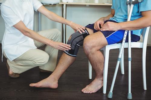
Post-surgical rehabilitation is a critical phase of recovery for individuals who have undergone surgical procedures. It encompasses a comprehensive approach to restoring physical function, mobility, strength, and overall well-being following surgery. One essential aspect of post-surgical rehabilitation is the management of scar tissue, which forms at the surgical incision site as part of the body’s natural healing process.
After surgery, the body initiates a complex cascade of biological processes to repair the injured tissues and restore normal function. One of the outcomes of this healing process is the formation of scar tissue at the site of the surgical incision. While scar tissue is essential for wound closure and tissue integrity, it can also pose challenges during the rehabilitation phase.
Purpose of Scar Management
The primary goals of scar management in post-surgical rehabilitation are to:
- Promote optimal scar healing: Enhance the appearance, texture, and flexibility of the scar tissue.
- Prevent complications: Minimize the risk of adverse outcomes such as hypertrophic scarring, keloid formation, or scar contractures.
- Improve functional outcomes: Enhance range of motion, strength, and mobility around the scar site to facilitate a return to normal activities and daily function.
Challenges of Scar Tissue
Scar tissue may present various challenges during post-surgical rehabilitation, including:
- Decreased flexibility: Scar tissue is often less elastic than normal skin, leading to stiffness and reduced range of motion, especially if it forms near joints or muscle attachments.
- Adhesions: Scar tissue may adhere to underlying structures such as muscles, tendons, or nerves, causing pain, discomfort, and restricted movement.
- Hypertrophic scars and keloids: Some individuals may develop raised, thickened scars known as hypertrophic scars or keloids, which can be cosmetically undesirable and may require additional interventions.
Understanding Scar Tissue
- Scar Formation: After surgery, scar tissue forms at the incision site as part of the body’s natural healing process. Initially, scars may appear red, raised, and firm, but they typically mature and become less noticeable over time.
- Scar Sensation: Scar tissue may be sensitive to touch, temperature changes, or movement, especially in the early stages of healing. This sensitivity usually diminishes as the scar matures.
Scar Management Strategies
- Early Intervention: Start scar management techniques once your surgeon gives approval, usually after the surgical wound has healed sufficiently. Early intervention can help minimize scar tissue buildup and improve the appearance and flexibility of the scar.
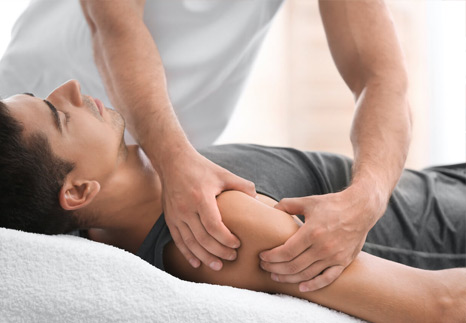
- Scar Massage: Gently massage the scar tissue using circular motions with clean fingertips or a soft cloth. Use a moisturizing lotion or silicone-based gel to reduce friction and promote scar tissue remodeling
- Compression Therapy: you may recommend wearing compression garments or dressings to apply gentle pressure to the scar. This helps flatten the scar and reduce swelling, redness, and discomfort.
- Silicone Products: Apply silicone sheets or gel to the scar as directed by your healthcare provider. Silicone has been shown to soften and flatten scars, reduce redness, and improve overall scar appearance.
- Sun Protection: Protect the scar from sun exposure by applying sunscreen with a high SPF or covering it with clothing when outdoors. Sun exposure can cause scars to darken and become more noticeable.
Scar Strength and Rehabilitation
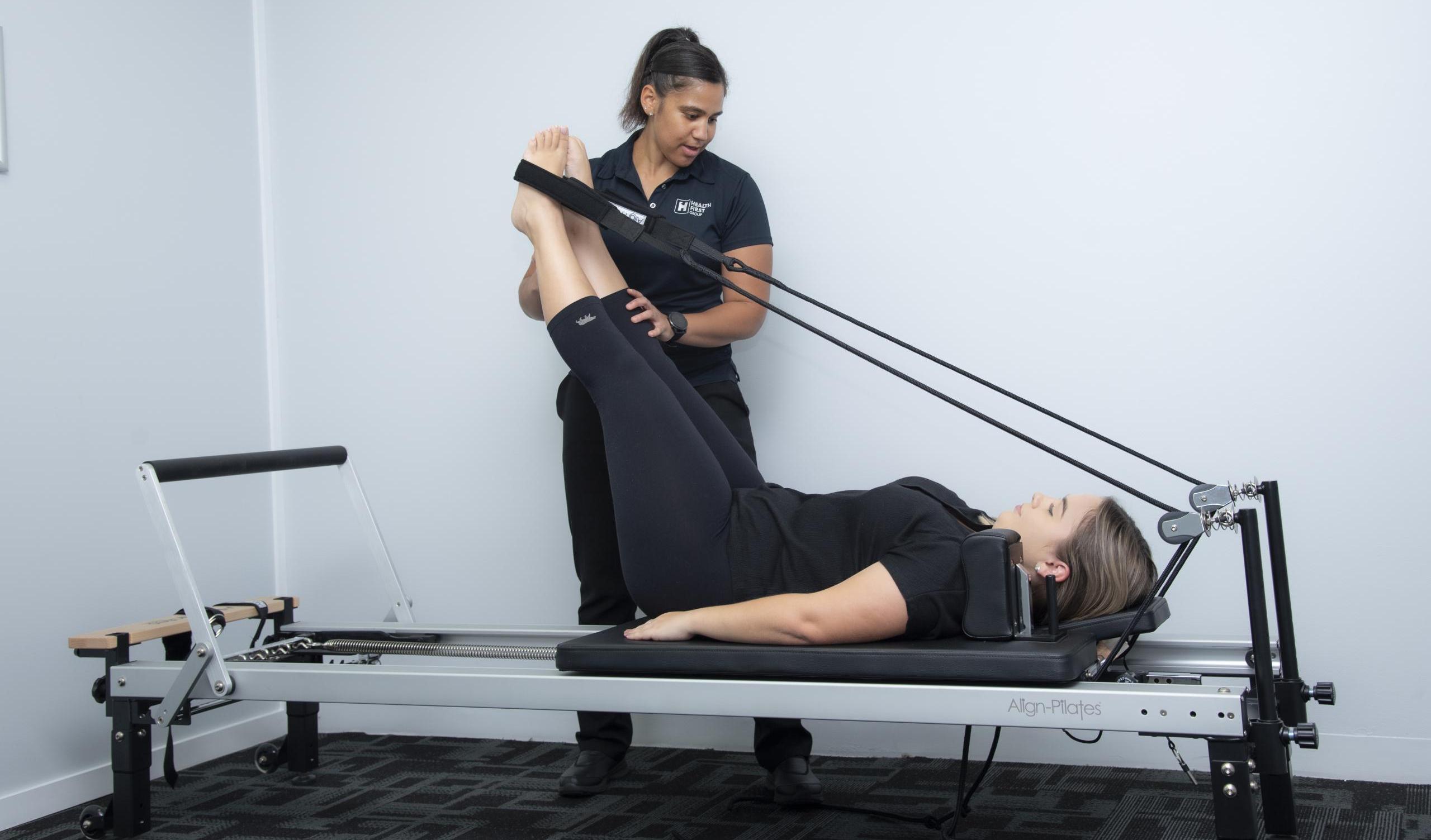
- Exercise Progression: Gradually incorporate gentle stretching and strengthening exercises into your rehabilitation program, taking care not to strain or stress the scar tissue excessively.
- Range of Motion: Perform range-of-motion exercises to improve flexibility around the scar site. Start with gentle movements and gradually increase the range as tolerated.
- Strengthening Exercises: Focus on exercises that target the muscles surrounding the surgical area to improve strength and stability. Begin with low-resistance exercises and gradually increase the intensity over time.
- Scar Protection: Avoid activities that place direct pressure or tension on the scar during the initial healing phase. Use padding or protective dressings if necessary to reduce friction and irritation.
Dry Needling
Dry needling involves inserting thin needles into trigger points within the scar tissue or surrounding muscles. These trigger points are areas of tightness and tenderness that can develop in response to injury or surgery. The needles stimulate the release of tension and promote the relaxation of overactive muscles, reducing pain and improving mobility. Dry needling can also enhance blood flow to the scar area, aiding in tissue healing and remodeling
Monitoring Scar Progression
- Regular Assessment: Keep track of the scar’s appearance, sensation, and overall healing progress. Report any changes or concerns to your surgeon during follow-up appointments.
- Scar Massage: Continue massaging the scar regularly to promote circulation, flexibility, and scar tissue remodeling. Monitor how the scar responds to massage and adjust your technique as needed.
Overall Rehabilitation
- Follow Rehab Plan: , performing home exercises, and following activity restrictions.
- Pain Management: Manage post-operative pain as directed by your surgeon using prescribed medications, ice packs, elevation, and relaxation techniques.
- Ultrasound Therapy: Ultrasound waves are used to penetrate deep into the tissues, promoting circulation and accelerating the healing process. This can help reduce scar formation and alleviate pain associated with scar tissue.
- Electrical Stimulation: Techniques like transcutaneous electrical nerve stimulation (TENS) or electrical muscle stimulation (EMS) can help manage pain and promote tissue healing.
- Rest and Recovery: Allow your body adequate time to rest and recover between rehabilitation sessions. Listen to your body and avoid overexertion or pushing through pain.
Conclusion
By incorporating these scar management and rehabilitation strategies into your post-surgical recovery plan, you can promote optimal healing, minimize scar-related complications, and improve overall functional outcomes.
“At Arunalaya Healthcare, we pride ourselves on being the best physiotherapy center in Delhi. Our dedicated team of experts offers top-notch physiotherapy treatment tailored to your needs. Experience the difference with the leading physiotherapy clinic in Delhi area. Our commitment to excellence ensures that you receive the best physiotherapy care possible. Trust Arunalaya Healthcare for the best physiotherapy treatment in Delhi. Our advanced physiotherapy solutions set us apart as the premier choice for your rehabilitation needs. When it comes to physiotherapy, our center stands out as the best in Delhi. Choose Arunalaya Healthcare for comprehensive physiotherapy solutions that deliver results. Visit Arunalaya Healthcare today and discover why we are the best physiotherapy center in Delhi.”
post-surgical rehabilitation | scar management techniques | managing scar tissue after surgery | dry needling for scar tissue | silicone sheets for scars | post-surgery recovery tips | scar massage benefits | range-of-motion exercises for scars | hypertrophic scars treatment | effective scar healing methods