Class 4 Laser Therapy

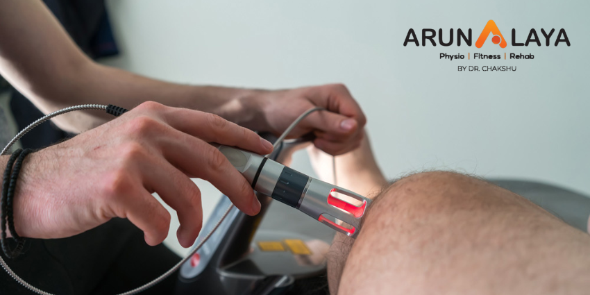
Your body has amazing systems that it can use to heal itself. However, sometimes it needs a little external help to get these systems fired up and working to your benefit. Class 4 laser therapy harnesses these processes using focused light. It stimulates a process called photobiomodulation (PBM).


Mechanism of action of class 4 laser:

  • The therapeutic effect of laser light is based on biostimulation and the acceleration of cellular processes that contribute to pain reduction and faster recovery after injury. The photomechanical wave stimulates nerve endings, which leads to pain relief. Biostimulation of the affected tissue increases oxygen uptake and improves blood circulation.
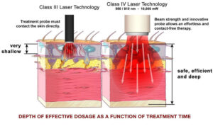
  • The unique combination of 30 W power and 1064 nm wavelength targets deep-lying tissues, maximises pain relief, and delivers strong thermic therapies within a short time.

Effects of class 4 laser therapy:

  • Reduced pain and swelling
  • Improved range of motion
  • To reduce pain and inflammation in musculoskeletal disorders and injuries and joint pain.
  • Increases tissue regeneration
  • Accelerates soft tissue and bone repair
  • Improve nerve regeneration & function as well as impact your cells to increase cell metabolism, enzymatic responses, collagen production, and promote angiogenesis
  • Improve your musculoskeletal health

What Are The Side Effects Of Laser Therapy?

  • Redness, swelling and itching. Treated skin may be itchy, swollen and red
  • Acne
  • Infection
  • Changes in skin color
  • Scarring
  • Turning of the eyelid

Rehabilitation of Hamstring Injuries

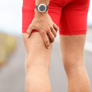
High-speed running, kicking, and sprinting frequently cause athletes to experience hamstring injuries, which primarily affect the muscle group known as the hamstrings.

To ensure successful rehabilitation for these injuries, it is important to adhere to the following general principles:

1. Train Movements and Muscles

The hamstrings, a muscle group consisting of three individual muscles, play different functional roles based on their anatomical positions. During sprinting, the biceps femoris (BF) undergoes the highest strain, the semitendinosus (ST) experiences the greatest lengthening velocities, and the semimembranosus (SM) primarily generates force. Therefore, it is important to target the injured muscle with specific exercises that aim to develop its particular functional role.

2. Prescribe Strength exercises to achieve a specific goal

Eccentric Training

The terminal swing phase of sprinting generates high eccentric forces. Modifying eccentric force deficits is essential as they contribute to the risk of future hamstring injuries. Hence, rehabilitation should prioritize training to develop the capacity for generating high eccentric force.

Get Long and Strong

The shortening of fascicles can predispose the hamstrings to (re-)injury, and eccentric training can help overcome this problem. Previous studies have demonstrated significant improvements in eccentric strength and fascicle lengthening with high-volume eccentric training programs incorporating the Nordic hamstring exercise. However, recent research has shown that similar improvements can be achieved with a low-volume program consisting of 2 sets of 4 repetitions once a week.

Isometric Training

During the swing phase’s conclusion, the hamstrings’ contractile element can remain relatively isometric, as the tendon primarily elongates the muscle-tendon unit. Maintaining a good isometric condition of the muscle reduces mechanical load and facilitates the tendon’s spring-like behavior during the stretch-shortening cycle.

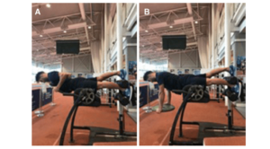
As hamstring injuries are consistently associated with fatigue, and most injuries occur in the final third of sprint training sessions, incorporating strength training under fatigued conditions has shown substantial reductions in injury rates. Isometric training of the hamstrings, such as utilizing the single-leg Roman chair hold, can enhance hamstring muscle endurance and serve as a valuable addition alongside the traditional Nordic hamstring exercises.

After an injury, the body’s response involves reducing myoelectric activity in the muscle as a protective mechanism to unload healing tissues. In some cases, this selective inhibition may persist and requires targeted rehabilitation. Isometric contractions have been shown to elicit higher voluntary muscle activation, making them effective in overcoming selective muscle inhibition. It is advisable to incorporate high-load isometric exercises to improve motor unit recruitment. In injuries where pain and disability are the primary concerns, isometric loading may be a more feasible approach before progressing to eccentric loading of the tissues.

Apply a multivariate model and target contributing factors to injury risk

Poor motor control of the pelvis can increase strain on the hamstrings and contribute to deficits in force production. Therefore, it is advisable to incorporate training for the lumbo-pelvic region, targeting movement in different planes.

Apart from the lumbo-pelvic region, the hip plays a vital role in maintaining optimal hamstring function. Weakness and decreased activation of the gluteus maximus serve as risk factors for hamstring injuries. Insufficient hip flexion exposes the hamstrings to a higher risk of injury, as the body compensates by increasing pelvic rotation during high-speed running, consequently placing greater strain on the hamstring muscles.

Cyclist’s Palsy

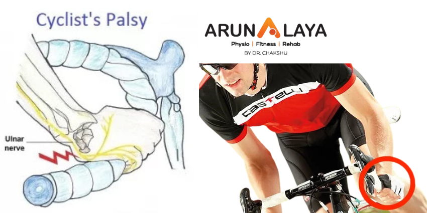
Cyclist’s palsy, also known by various names such as handlebar palsy, ulnar tunnel syndrome, ulnar nerve compression, Guyon Canal Syndrome (GCS), bicycler’s neuropathy, or tardy ulnar palsy, is an overuse injury that primarily affects the hands and fingers. It occurs when the nerves in the wrist or the side of the palm near the pinky finger become compressed due to repetitive stress.

Cause of cyclist palsy

  • Cyclist’s palsy can cause both motor and sensory symptoms.
  • The motor symptoms can include weak hand grip and difficulty using fingers for precise tasks.
  • Whereas the common sensory effects include numbness, tingling, and pain.

Although cyclist’s palsy is increasingly common, it is often underreported among cyclists. In fact, a study revealed that 7 out of 10 participants reported experiencing motor or sensory symptoms. Severe nerve injury can lead to paralysis or irreversible loss of sensation in the affected hand.

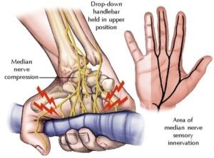
The pressure exerted on the handlebars during prolonged cycling can irritate the nerves in the palm. The highest pressure occurs where the median and ulnar nerves enter the hand, which corresponds to positions like “tops,” “ramps,” “hoods,” and “drops.” The “drops” position applies the most pressure on the ulnar nerve, while the “hoods” position applies slightly less pressure. The “tops” position places significant pressure on the palm at the base of the ring finger. The “drops” position can also cause excessive wrist extension, increasing pressure on the carpal tunnel. If a cyclist already has nerve compression at the neck or elbow, it can be more easily triggered at the palm, potentially leading to carpal tunnel syndrome or cubital tunnel syndrome.

Signs and Symptoms

  • include numbness, tingling, and sensory changes in the little finger and the ring finger on the side closest to the little finger
  • the palm in that area may also become numb, while there is no numbness on the back of the hand.
  • The symptoms can vary depending on the location of pressure. Sometimes manifesting as numbness or weakness, or a combination of both.
  • When the median nerve is affected, numbness and tingling occur on the palm side of the thumb, index, long, and ring fingers (on the side closest to the middle finger). But there is no numbness on the back of the hand.
  • Prolonged or severe pressure on the nerves can also weaken the associated muscles. Some cyclists may experience pain along with hand numbness.

Treatment

Limiting cycling is the most effective treatment for cyclist’s palsies. However, there are other measures that can allow cyclists to continue their activity while reducing the risk of exacerbating the condition. These include :

  • Limiting the length or distance of the ride
  • Having enough rest between longer cycling sessions
  • Changing positions of grip on the handlebars
  • Changing to a transverse handlebar
  • Adjusting the seat height
  • Using gloves to reduce or distribute pressure. The pressure can be reduced with foam or gel padding in the palm of the glove.

Top 3 Cyclist Palsy Exercises

In addition to these measures, exercises play a crucial role in long-term recovery and preventing recurrence of cyclist’s palsy. These exercises primarily focus on strengthening the muscles, ligaments, and tendons in the hands. The top three recommended exercises for cyclist’s palsy are as follows:

  1. Finger bending exercise: Begin by stretching your hand and then bend the fingers of the affected hand at a right angle, holding them in that position for approximately 10 seconds. Ensure that your fingers remain straight during the exercise. Repeat this process five times.
  2. Finger squeeze: Take a small object like a coin or a sheet of paper and squeeze it between two fingers, holding the grip for 10 seconds. Repeat this exercise five times for each set of fingers.
  3. Grip strengthening exercise: This exercise targets a weak hand grip. Squeeze a rubber ball with the affected hand and hold for 10 seconds and then release. Repeat 10 times, and that’s one set. Aim for 3 sets of 10 as you gradually build up grip strength.


Low Back Pain Relief

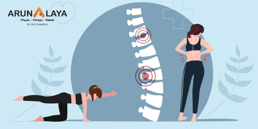
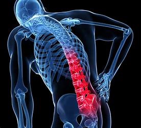
Most types of low back pain are often referred to physical therapy as one of the first-line treatments. Physical therapy for low back pain includes guided therapeutic exercises that strengthen the lower back muscles and condition the spinal tissues and joints.

The short- and long-term goals of physical therapy for back pain typically include the following:

  • Decrease painful symptoms in the lower back and/or leg
  • Improve low back function to tolerate daily activities as independently as possible
  • Increase the spine’s flexibility and improve its range of motion
  • Formulate a maintenance program to prevent the recurrence of back problems

The exercises are intended to provide flexibility and strength training to the entire kinetic chain—groups of body segments, joints, and muscles that work together to perform bodily movements.

Physical therapy helps restore the patient’s ability to perform daily activities with little-to-no discomfort. Large-scale studies have shown that physical therapy can provide up to 60% improvement in lower back pain and other symptoms.

Four Categories of Therapeutic Exercises for Back Pain

Therapeutic exercises are typically performed or learned under the guidance of a physical therapist and use a combination of the approaches described below.

  1. Core-Strengthening Exercises

In simple terms, the body’s core can be described as the area surrounded by the stomach muscles in front, the spinal and buttock muscles at the back, the bottom of the lungs or the diaphragm on top, and the muscles of the pelvis and hip at the bottom.  A weak core distributes weight unequally throughout the spine and into the legs, causing back pain or worsening existing back pain. Core-strengthening exercises help build strength and endurance in the core muscles, significantly reducing pain and improving functionality in the lower back.

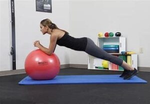
Easy-to-perform core-strengthening exercises to relieve back pain include the pelvic tilt, cat-cow pose, bird dog, high and low planks, crunches, and exercises performed using a Swiss Ball.

  1. Lumbar Stabilizing Exercises

A strong spine needs strong hips and legs to support the lower body, and to walk, bend, and twist effortlessly. Inefficiency of the muscles in the hips and legs has been proven to cause spinal instability and pain. Stretching exercises can activate and strengthen these muscles, such as the iliopsoas and hamstrings; improve coordination between the hip and spine; and aid in the transfer of forces across the lower back, pelvis, and legs.

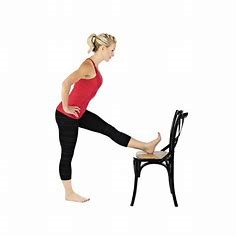
Hip and leg exercises for beginners with lower back pain typically include hamstring stretches, squats, downward dog, planks with leg lifts, and lunges.

  1. Aerobic exercises

Aerobic or cardiovascular exercise not only helps maintain a healthy heart but also heals the spinal muscles. Cardiovascular exercise involves synchronized movement in the body, which increases the heart rate, thereby improving circulation, oxygen content within the cells, and energy production in the tissues. Through these mechanisms, the painful spinal muscles respond by becoming less stiff and more mobile.

Simple aerobic exercises for back pain include brisk walking, a stationary bike, and an elliptical trainer. Low-impact aerobic conditioning can be achieved through aquatic exercise. The buoyancy of water supports the body’s weight, reducing stress on the spine and allowing for a greater range of motion.

  1. Postural Training

Supported posture minimizes strain on the body by maintaining a balance of the muscles and bones.  Unsupported posture can result from habit, painful symptoms, or ergonomics at work or home. Specifically in the spine, using incorrect posture can limit the movement of the tendons and muscles, making regular day-to-day movements difficult and painful.  Posture correction exercises aim to stretch and strengthen the back and abdominal muscles and the kinetic chain, which help stabilize the spine.

Common posture correction exercises include calf stretching, seated squats, pelvic tilts, and abdominal strengthening exercises.

Physical therapy is a multi-component program that includes education, training, strength, flexibility, and endurance. A physical therapist plays a key role in designing a program suitable to the needs and limitations of the patient. A therapist’s goal is to teach the proper exercise technique, so the patient can perform the exercise on their own.

Duration of a Physical Therapy Program for Low Back Pain

The total length of an exercise program depends on the severity and duration of the low back symptoms.

  • For chronic low back pain, physical therapy typically starts with an 8-week program performed under the guidance of a physical therapist.
  • A longer-term maintenance or rehabilitation therapy is prescribed after the guided phase, which can be done at home.

The duration of the guided program may increase or decrease based on the outcome of the treatment.

Physical therapy may be combined with other nonsurgical treatments for the lower back to improve the overall pain outcome. Hands-on alternative treatments, such as massage therapy, manual therapy, and accupuncture may be beneficial when performed in combination with a physical therapy regimen.

ROLE OF PHYSIOTHERAPY IN HIP DYSPLASIA

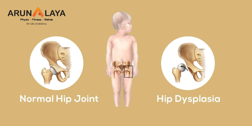
Hip dysplasia or developmental dislocation of the hip (DDH), is an abnormal formation of the hip joint. In hip dysplasia, the femoral head (the ball) fits too loosely into the acetabulum (the socket), causing instability. The disorder typically develops during gestation or shortly after birth, but may develop later in childhood. In the past, this condition was referred to as congenital dysplasia of the hip (CDH).

CAUSES

Many people are born with hip dysplasia but are likely unaware because symptoms don’t often present themselves until adulthood.

Other times, it is typically developed within the first year of a person’s life.

Adults can develop hip dysplasia, but it is most often a diagnosis that wasn’t determined in childhood likely from a lack of symptoms.

PHYSICAL SYMPTOMS

Someone suffering from hip dysplasia may:

  • Have legs that are different lengths
  • Have a leg that turns outward
  • Have uneven skin folds on their groin or thigh
  • Lean to one side when standing
  • Have minimal or complete loss of range of motion in the hip
  • Limp when walking

INTERNAL SYMPTOMS

Someone suffering from hip dysplasia may have internal symptoms like:

  • Pain when sleeping on the affected hip
  • Pain in the groin that increases with activity
  • A sensation of popping, snapping or catching at the hip
  • A stiff hip joint

PHYSIOTHERAPY TREATMENT

Weight loss and other lifestyle changes can improve the condition.

Physiotherapy includes

  • leg stretching particularly hip abductions

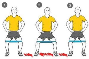
  • Banded lateral walk

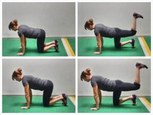
  • Glute activation

When engaging in physical therapy exercises, it is usual for patients to be provided with applied heat before the exercise regimen and ice packs afterwards.

Physical therapy is a great option for patients to help:

  • Strengthen muscles surrounding the hip
  • Joint mobility
  • Correct poor posture
  • Tendon inflammation
  • Gait
  • Body awareness

Advance physiotherapy treatment

  • MFR
  • Kinesiology tape
  • Dry needling

Medical professionals also may recommend:

  • Including low- or non-impact exercises into your weekly routine to strengthen muscles and increase range of motion like:
  • Swimming
  • Aquatic therapy
  • Cycling
  • Bodyweight exercises
  • Losing or maintaining weight to reduce the stress and pain in the hip
  • Hippotherapy to improve motor function

Shock Wave Therapy: How it Works?

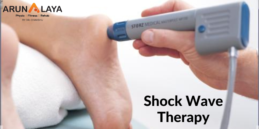
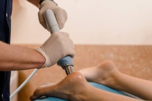
Shock Wave therapy is a technique that uses high-energy sound waves rather than electrical shocks to aid in the healing process following an injury.

SWT is a cutting-edge treatment that uses acoustic shock waves to break up soft tissue calcifications, boost collagen production, release growth factors, and accelerate your body’s healing process in order to reduce pain and keep you active.

It’s a lesser-known physiotherapy technique, yet it can be beneficial.

Injuries that can be treated

Shock wave therapy is used to treat musculoskeletal injuries, particularly those areas involving connective tissue and bone.
The following are some examples of common injuries treated with shockwave therapy:

  • tennis/golf elbow
  • rotator cuff tendonitis
  • calcification
  • stress fractures
  • trochanteric bursitis in the hip
  • patellar tendonitis
  • jumper’s knee
  • hell spurs
  • Achilles tendonitis
  • Morton’s neuroma in the foot

Shock wave therapy treatment has been shown to be an excellent approach to start the body’s healing process and reduce discomfort while regaining movement.
Most people require only three treatments per week.

This therapy improves blood circulation and hastens the repair of chronic musculoskeletal disorders in the bones, tendons, ligaments, and muscle.

How does it work?

  • Shockwave treatment stimulates self-healing by using radial acoustic shockwaves.
  • These shockwaves, which are not electrical but rather sound shockwaves, are administered through a special wand hand piece that dispenses the waves for a brief amount of time directly to the affected injured tissue areas.
  • The pressure from the shockwaves will be transferred to the tissue. As a result, special micro-cavitation bubbles form, expand, burst, and produce a force known as a resultant force.
  •  This passes through the tissue and stimulates the cells responsible for connective tissue and bone mending, so activating the body’s inherent self-healing mechanism.
  • These processes involve increased metabolism and blood circulation. It means that your damaged tissue receives better healing treatment from your body, and the damaged tissue will begin to regenerate itself, with an accompanying inflammatory response allowing the healing process to take effect.
  • Some people find it slightly uncomfortable to go through. However, these people are frequently in the minority, and the majority are able to handle the five-minute treatments with ease.
  • Though there will be no pain immediately following the treatment, there is a potential of discomfort in the hours that follow.
  • This discomfort can last for up to two days, therefore patients should limit their physical activity and avoid using anti-inflammatory drugs or ice.

How does it feel?

ESWT hurts, and the degree of pain varies from person to person. The majority of patients perceive it as slightly uncomfortable but not painful.
It also affects where we treat you. Body portions with less skin and muscle, such as above the hip bone, can be more sensitive than meatier areas, such as the calves.

OSTEOPOROSIS MANAGEMENT IN PHYSIOTHERAPY

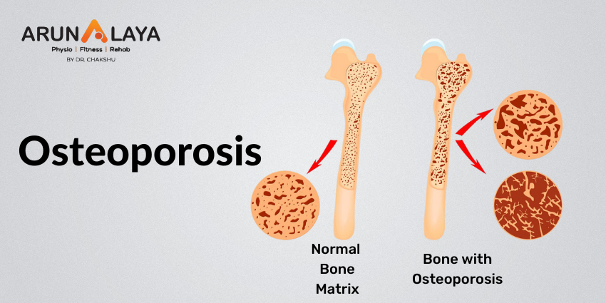
Osteoporosis is a condition in which the bones in the body become weak and brittle as a result of tissue loss over time. This illness is most common in the elderly, but it can also occur in middle-aged patients.

Although this condition can affect any bone in the body, it most commonly affects the following:

  • Spine
  • Hips
  • Waist

Because of bone fragility, people with osteoporosis are susceptible to fractures.

Causes

While weak bone structures are caused by ageing factors, smoking, and excessive alcohol use, a prolonged sedentary lifestyle can also play a role. Cigarettes contain toxic substances that interfere with the bones’ natural ability to repair, while alcohol interferes with calcium absorption.

Habitual physical inactivity promotes the activity of osteoclasts (the cells responsible for bone resorption).

Another factor that contributes to osteoporosis is hormonal change associated with ageing. Women going through menopause may be more likely to develop osteoporosis later in life due to lower oestrogen levels (a hormone that helps manufacture bone mass).

Anyone can get osteoporosis, and the risk rises with age.

Role of Physiotherapy

Physiotherapy can aid in the prevention and management of osteoporosis.
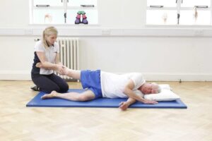
Over the course of weeks and months, a physical therapist will design a personalised training programme to strengthen your bones and muscles.
This improves your balance and reduces your chances of falling.

Physical therapy can also aid in the recovery from an osteoporosis-related accident.

Physical therapy often involves performing stretches or exercises, usually done in repetition or sets. But physical therapists also use a variety of other techniques to improve movement.

These include:

  • Manual therapy
  • Kinesiology taping
  • Dry needling
  • suggestions for lifestyle changes
  • MFR
  • heat or cold therapy
  • ultrasound / electrical stimulation
  • Stability training

Benefits of PT

  • Physiotherapy treatments aid in the strengthening of bones and muscles
  • Regular and targeted exercise is essential for the prevention and management of osteoporosis.
  • Working with a physiotherapist can help you with the following:
    1. improve your balance
    2. lower your chances of falling
    3. stretch and strengthen your muscles
    4. correct stooped posture
    5. prevent further bone loss
    6. reduce pain

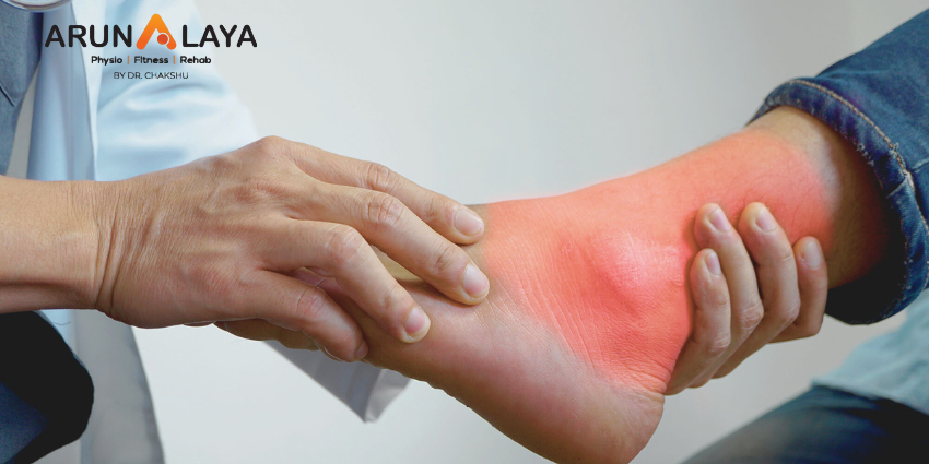
Understanding Lateral Ligament Injury of the Ankle

Lateral ligament injury is among the most prevalent types of sports injuries addressed by physiotherapists. Men and women are estimated to suffer from lateral ankle sprains at roughly the same rates.

A lateral ligament injury of the ankle is a common injury that occurs when the ligaments on the outer side of the ankle are damaged. The most frequently affected ligaments are the anterior talofibular ligament (ATFL), the calcaneofibular ligament (CFL), and the posterior talofibular ligament (PTFL). These ligaments are responsible for providing stability to the ankle joint.

Lateral ankle sprains are also known as inversion or supination ankle sprains. The complex of ligaments on the lateral side of the ankle is torn by varied degrees as a result of a forced plantarflexion/inversion movement.

Individuals who sustain multiple recurrent ankle sprains have been documented to have functional and mechanical instability, as well as an increased risk of re-injury. Small fractures surrounding the ankle and foot (e.g. Pott’s fracture) and straining or rupture of the muscles around the ankle (e.g. calf, peroneii, tibialis anterior) should also be avoided even if they are the less prevalent causes of ankle pain.

Lateral Ankle Ligament Tear Types:

Lateral ligament injuries of the ankle can be classified into three main types based on the ligaments involved and the severity of the tear:

Anterior talofibular ligament (ATFL) injury: This is the most common type of lateral ligament injury and often occurs in isolation. It involves the stretching or tearing of the ATFL, which connects the talus bone to the fibula. It is commonly associated with ankle inversion injuries.

Calcaneofibular ligament (CFL) injury: The CFL is located slightly below the ATFL and connects the calcaneus (heel bone) to the fibula. CFL injuries typically occur along with ATFL injuries or in more severe sprains. In some cases, the ATFL and CFL can be injured together, resulting in a higher-grade ligament tear.

Posterior talofibular ligament (PTFL) injury: The PTFL is the least commonly injured ligament in lateral ligament injuries of the ankle. It connects the talus bone to the fibula at the back of the ankle. PTFL injuries usually occur in severe ankle sprains or high-energy trauma.

CAUSES

Ankle sprain: The most common cause of a lateral ligament injury is an ankle sprain, which typically occurs when the foot rolls inward, resulting in stretching or tearing of the ligaments on the outer side of the ankle.

Sports injuries: Activities that involve quick changes in direction, jumping, or running on uneven surfaces increase the risk of ankle sprains and ligament injuries.

Trauma: Direct trauma or impact to the ankle joint, such as a fall or a collision, can cause damage to the lateral ligaments.

CLINICAL FEATURES

  • Pain on the outer side of the ankle
  • Swelling and bruising
  • Difficulty walking or bearing weight on the affected foot
  • Instability or a feeling of the ankle giving way
  • Limited range of motion in the ankle joint.

RISK FACTORS

Body mass index, slow eccentric inversion strength, quick concentric plantar flexion strength, passive inversion joint position sense, and peroneus brevis reaction time were all linked to an elevated risk of lateral ankle injury.

ASSESSMENT OF ANKLE JOINT

  • Amount of instability present by assessing the grade of the sprain;
  • Loss of Range of motion (ROM);
  • Loss of the muscle strength;
  • Level of reduced Proprioception.

OBSERVATION

Any symptoms of injury, inflammation, skin colour changes, or muscle atrophy or hypertrophy are noted. Following that, the foot and ankle are observed in two separate positions: non-weight bearing and weight bearing. Take note of the gait pattern, degree of limp (if any), and facial expression when bearing weight.

HISTORY

Mechanism of injury: A plantarflexion/inversion injury would indicate damage to the lateral ligament, whereas a dorsiflexion/eversion injury would indicate damage to the medial ligament. Previous history of injury on the same side will give clues as to whether the ankle was unstable to begin with, or that a previous injury wasn’t properly rehabilitated.

History of injury on the other side as well may indicate a biomechanical predisposition towards ankle injuries.

GRADES

Grade 1: Mild sprain with minimal stretching or tearing of the ligament fibers. There may be slight swelling, tenderness, and minimal loss of function.

Grade 2: Moderate sprain with partial tearing of the ligament fibers. This grade is characterized by increased swelling, bruising, pain, and difficulty walking or bearing weight on the affected ankle.

Grade 3: Severe sprain with complete tearing or rupture of the ligament. Significant swelling, bruising, pain, and instability are commonly observed. Walking or weight-bearing may be extremely difficult or impossible.

SPECIAL TEST

An anterior draw is performed to assess the ATFL and CFL integrity. The heel is grabbed with the ankle in plantarflexion, and the tibia is stabilised and dragged anteriorly.
The talar tilt is used to evaluate the integrity of the ATFL and CFL laterally, as well as the deltoid ligament medially. The heel is grabbed again, the tibia is stabilised, and the talus and calcaneus are pushed laterally and medially.
Beginning with a simple single leg stance, proprioception can be measured in a variety of more challenging methods. The patient can do it on the normal side first to give the therapist a sense of what is typical, and then try it on the injured side.

DIFFERENTIAL DIAGNOSIS

  • Ankle fracture (medial/lateral malleolus, distal tibia/fibular)
  • Damage to the medial ligament
  • Dislocated ankle
  • Other soft tissue damage (peroneal tendons, muscle strain)

TREATMENT

REDUCE PAIN AND SWELLING

The RICE regimen (Rest, Ice, Compression, and Elevation) can be used to minimise pain and swelling in the first 48-72 hours after an acute lateral ligament damage.

If weight bearing is too painful, the patient can use elbow crutches for 24 hours and remain non-weight bearing. However, it is critical to begin at least partial weight bearing as soon as possible, along with a regular heel-toe gait pattern, since this will help to reduce pain and swelling.

Gentle soft tissue massage and light stretches can be conducted to help with the clearance of oedema, as long as they are painless.

Range of motion exercises: These exercises aim to restore the normal range of motion in the ankle joint and may involve gentle ankle rotations, ankle pumps, and alphabet exercises.

Strengthening exercises: Strengthening the muscles around the ankle joint helps provide support and stability. Common exercises include calf raises, toe raises, ankle inversions and eversions (using resistance bands or manual resistance), and single-leg balance exercises.

Proprioceptive and balance exercises: These exercises improve the body’s awareness of joint position and enhance balance and stability. Examples include standing on one leg, balance board exercises, and wobble board exercises.

RETURN TO FUNCTIONAL ACTIVITY

  • Twisting
  • Jumping
  • Hopping on one leg
  • Running
  • Figure of 8 running

Before returning to full functional activity the patient should have full range of pain free movement in the ankle, normal strength and normal proprioception. If returning to sports, the athlete should be encouraged to wear an ankle brace or to tape the ankle for a further 6 months to provide external support.

Role of Physiotherapy in Breast Cancer Recovery

Treatment to the chest and axilla for breast cancer can lead to secondary complications in the arm like pain, reduced movement of the shoulder joint, muscle weakness, swelling (lymphedema) and difficulty to carry out activities of daily activities. The problems can persist for many years after treatment. Guidelines state that when indicated patients with breast cancer should be referred to physiotherapy early, however this is not a routine practice.
There is a need for a proactive model of care which encourages early exercise-based rehabilitation.

Read More

Tingling and Numbness

Tingling and Numbness

Sometimes we can feel a sensation of pins and needles pricking us in any part of the body. In medical terms, this is known as Paresthesia. People generally notice these sensations in hands, feet, arms, and legs. If numbness and tingling persist and there’s no obvious cause for the sensations, it could be a symptom of a disease or injury, such as multiple sclerosis or carpal tunnel syndrome.

Read More