Kinesiology tape

Kinesiology tape

Introduction

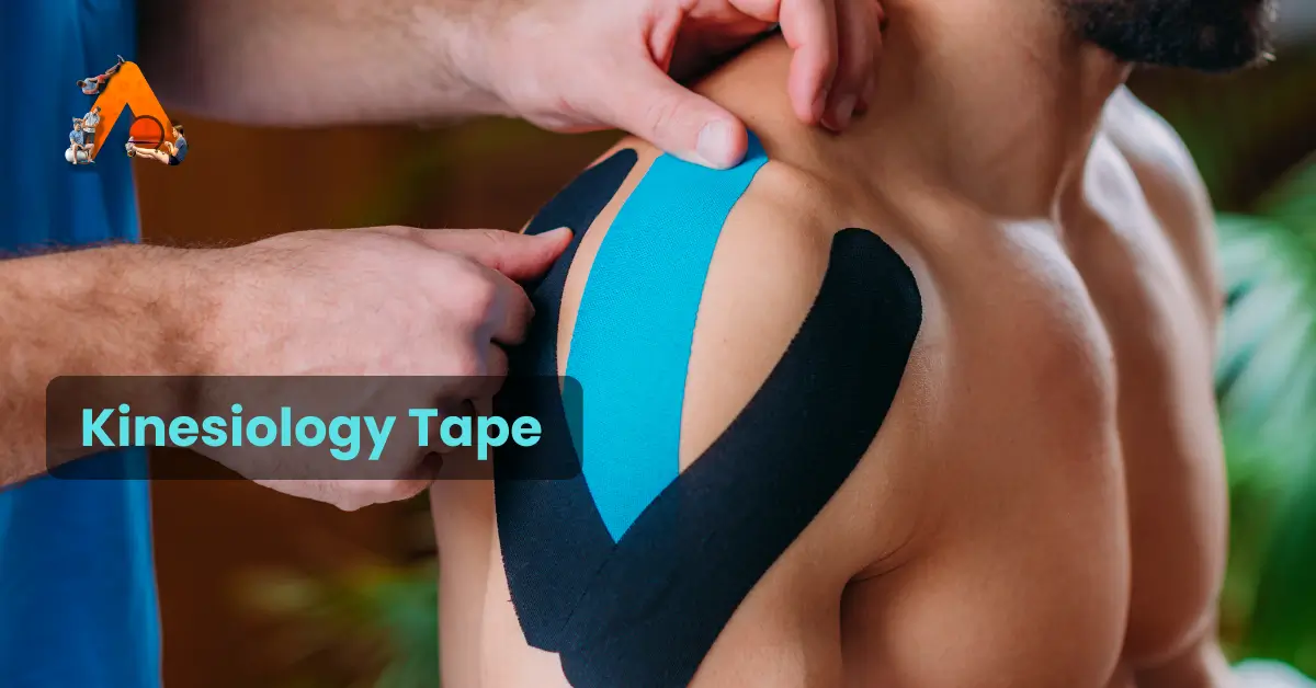
Kinesiology tape has become a widely recognized tool in physical therapy and sports medicine, praised for its ability to support muscles and joints, alleviate pain, and enhance physical performance. Developed by Dr. Kenzo Kase in the 1970s, this innovative tape mimics the skin’s elasticity, enabling natural movement while providing therapeutic benefits. Its diverse applications, ranging from injury recovery to chronic pain management, have made it a staple in rehabilitation programs worldwide.

Key Features of Kinesiology Tape

  1. Material Composition
    Kinesiology tape is crafted from a blend of cotton and nylon, ensuring breathability and stretchiness. It adheres to the skin using a strong, water-resistant adhesive and is often available in latex-free, hypoallergenic options to suit sensitive users.
  2. Functionality
    By creating a gentle lift in the skin, kinesiology tape helps reduce pressure on underlying tissues. This unique lifting mechanism improves circulation and decreases pain signals sent to the brain, promoting faster recovery.
  3. Application Techniques
    The tape can be applied in various patterns—such as I, Y, or X—depending on the user’s needs. Proper application is essential to achieve desired outcomes, and consulting a trained professional is often recommended for optimal results.

Benefits of Kinesiology Tape

  • Pain Relief
    Many users report decreased pain after application, as kinesiology tape interrupts pain signals and fosters a sense of relief.
  • Reduced Inflammation
    Enhanced blood flow and lymphatic drainage help mitigate swelling and inflammation in injured areas.
  • Enhanced Performance
    Athletes use kinesiology tape to support fatigued or injured muscles, enabling better performance during competition.
  • Muscle Support
    The tape stabilizes weak or injured muscles without hindering movement, facilitating improved functionality during physical activity.
  • Posture Correction
    By offering feedback through the skin, kinesiology tape aids in correcting postural issues, increasing awareness of body positioning.
  • Support for Chronic Conditions
    Its role in conditions involving muscle overuse or chronic pain, such as tendonitis or sprains, is particularly noteworthy.

Specific Conditions Treated by Kinesiology Tape

  1. Sports Injuries
    Kinesiology tape is commonly applied for injuries like ankle sprains, shoulder impingement, and tennis elbow. It supports the injured area while allowing continued movement, aiding recovery.
  2. Chronic Pain
    Conditions such as fibromyalgia and chronic low back pain benefit from the tape’s ability to stabilize affected areas and reduce discomfort.
  3. Muscle Strains and Sprains
    Acute muscle strains and sprains are managed effectively using kinesiology tape, which alleviates pain by reducing pressure on sensitive tissues.
  4. Postural Issues
    Poor posture, a common cause of pain, can be corrected with kinesiology tape, which encourages proper alignment of the spine and shoulders.
  5. Swelling and Inflammation
    The tape’s ability to improve lymphatic drainage and circulation makes it effective in reducing swelling from conditions like lymphedema or post-surgical recovery.
  6. Joint Pain
    Arthritis and general joint pain can benefit from kinesiology tape’s supportive properties, which stabilize joints without limiting mobility.
  7. Post-Surgical Rehabilitation
    During rehabilitation after surgery, kinesiology tape helps restore mobility and function while protecting healing tissues.
  8. Trigger Points
    By lifting the skin over tense muscles, kinesiology tape relieves pain from trigger points, reducing tension and improving flexibility.

Effectiveness of Kinesiology Tape for Chronic Pain

  1. Pain Relief
    Research suggests kinesiology tape can provide significant pain relief, especially when compared to minimal intervention. A meta-analysis demonstrated notable reductions in pain scores among users treated with kinesiology tape.

2. Mechanisms of Action

  • Decompression of Tissues: The tape’s skin-lifting effect reduces pressure on tissues, altering sensory signals and potentially decreasing pain perception.
  • Improved Circulation: Enhanced blood and lymphatic flow reduces inflammation and promotes healing, particularly in conditions involving swelling.

3. Support for Muscle Function
Kinesiology tape stabilizes muscles and joints, improving function and reducing discomfort without restricting movement.

Considerations and Limitations

  1. Short-Term vs. Long-Term Effects
    While kinesiology tape provides short-term relief, its long-term effectiveness is uncertain. For instance, pain relief observed with taping may not persist six months post-treatment.
  2. Multidisciplinary Approach
    Kinesiology tape is often used alongside other therapies like manual therapy and exercise. This combined approach ensures a more comprehensive treatment for chronic pain or injury recovery.

Conclusion

Kinesiology tape is a versatile tool in managing pain, supporting recovery, and enhancing physical performance. Its ability to improve circulation, provide muscle support, and promote healing makes it a valuable addition to rehabilitation programs. However, it should not be seen as a standalone solution but rather as part of a holistic treatment plan. Consulting healthcare professionals ensures proper application and optimal outcomes, making kinesiology tape an essential component of modern therapeutic practices.

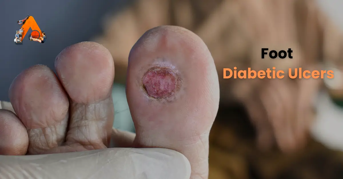
Diabetic Ulcers

Physiotherapy Management of Diabetic Ulcers

Diabetic ulcers are a significant complication of diabetes, affecting approximately 15% of individuals with the condition. This blog provides an overview of diabetic ulcers, including their types, causes, risk factors, clinical features, assessment, and physiotherapy management.

Introduction

A diabetic ulcer is an open sore or wound commonly located on the bottom of the foot. These ulcers arise from factors such as poor circulation, high blood sugar levels, and nerve damage. The condition is further complicated by delayed healing due to restricted blood supply and infections. Understanding the types, assessment, and management of diabetic ulcers is crucial for effective treatment and prevention.

Types of Diabetic Ulcers

Neuropathic Ulcers

  • Occur due to peripheral diabetic neuropathy without ischemia caused by peripheral artery disease.

Ischemic Ulcers

  • Arise where there is peripheral artery disease without diabetic neuropathy involvement.

Neuro-ischemic Ulcers

  • Result from a combination of peripheral neuropathy and ischemia.

Nature and Sites

  • Diabetic ulcers are deep and spreading, commonly affecting the toes, soles of the feet, and occasionally, the legs, hands, fingers, and arms.

Diabetic Foot Ulcers

Diabetic foot ulcers are full-thickness wounds on the plantar surface of the foot. They are characterized by neuropathy, ischemia, and infection. High plantar pressures often cause these ulcers, especially at the second metatarsal head.

Etiology of Diabetic Ulcers

Diabetes affects nerves through two primary pathways:

  1. Free Radical Damage
    • Rogue oxygen molecules damage cells, leading to inflammation.
  2. Reduced Vascular Supply
    • Narrowing of arteries restricts blood flow, delaying wound healing and causing nerve damage.

High blood sugar levels and associated conditions (e.g., high cholesterol, hypertension) exacerbate these effects, leading to neuropathy, impaired sensation, muscle weakness, and abnormal foot structures.

Risk Factors for Diabetic Foot Ulcers

  • Diabetes
  • High cholesterol levels
  • Coronary heart disease
  • Hypertension
  • Atherosclerosis
  • Previous history of foot ulcers or amputations
  • Poor glycemic control
  • Smoking

Classification of Diabetic Foot Ulcers

The Meggitt-Wagner Classification outlines the severity of diabetic ulcers:

  • Grade 0: No ulcer in a high-risk foot.
  • Grade 1: Superficial ulcer without underlying tissue involvement.
  • Grade 2: Deep ulcer penetrating to ligaments or muscle, no bone involvement.
  • Grade 3: Deep ulcer with cellulitis, abscess, or osteomyelitis.
  • Grade 4: Localized gangrene.
  • Grade 5: Extensive gangrene involving the whole foot.

Clinical Features of Diabetic Ulcers

  • Redness, swelling, or discoloration around the wound.
  • Break in the skin, discharge, or itching.
  • Dryness and pain.
  • Thickened or callused skin around the ulcer.
  • Fever and chills in advanced stages.

Assessment of Diabetic Ulcers

History Taking

  • Ulcer onset, self-treatments, history of infection, and prior interventions.
  • Medical history of diabetes, autoimmune, metabolic, or cardiac conditions.
  • Surgical history, including previous amputations or debridement.
  • Personal history, such as smoking or alcohol use.

Examination

  • Skin Status: Visual inspection for peeling, maceration, and fissures.
  • Neurological Status: Semmes-Weinstein monofilaments or 128 Hz tuning fork to assess sensation.
  • Vascular Status: Checking pedal pulses, capillary refill time, and Doppler ultrasound findings.
  • Ulcer Evaluation: Noting site, size, depth, discharge, and surrounding tissue condition.

Imaging

  • X-rays for deep wounds and MRI for detecting osteomyelitis or abscesses.

Physiotherapy Management

Physical Therapy Modalities

  1. Thermotherapy
    • Infrared lamps and ultrasound therapy improve blood flow and cell metabolism.
  2. Electrotherapy
    • Electrical stimulation enhances wound repair.
  3. Shockwave Therapy
    • Focuses sound waves to improve soft tissue healing.
  4. Laser Treatment
    • Effective for microcirculation and pain relief.
  5. Galvanic Current
    • Direct electrical current for tissue repair.

Exercises

  • Range of motion, stretching, Buerger-Allen, and proprioception exercises enhance blood flow and sensory input.

Shoe Modifications

  • Special shoes with rocker bars, plastazote insoles, and proper cushioning distribute pressure evenly and reduce injury risks.

Nutrition Advice

  • Adequate intake of iron, vitamins (A, B12, C), zinc, and proteins is essential for wound healing.

Prevention of Diabetic Ulcers

Primary Prevention

  • Improved glycemic control and reduced cardiovascular risk factors.
  • Routine foot examinations for early detection.

Secondary Prevention

  • Quit smoking and avoid activities that may injure the feet.
  • Daily foot inspections and proper hygiene.
  • Use appropriate footwear and moisturizers (avoiding application between toes).

Conclusion

Diabetic ulcers are a complex yet preventable complication of diabetes. With appropriate physiotherapy modalities, shoe modifications, and preventive measures, patients can achieve improved outcomes and a better quality of life. Early intervention and comprehensive care play a vital role in minimizing the risks associated with diabetic ulcers.

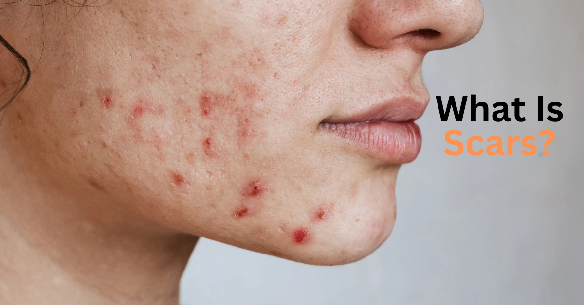
What is scars?

What are scars?

Introduction

Scars form as a natural part of the body’s healing process after skin damage. When the skin sustains an injury, it repairs itself by generating new tissue to close the wound and fill in any gaps created by the injury. This tissue, known as scar tissue, is primarily composed of collagen. The appearance of a scar depends on various factors such as the type of injury, the location of the wound, treatment methods, and even genetic factors.

What is Scar Tissue?

Scar tissue is a collection of cells and collagen that covers the site of an injury. Unlike normal skin, scar tissue has distinctive characteristics due to its unique structural makeup

Factors Influencing Scar Appearance

  • Cause of injury: Such as surgery, burns, or severe acne
  • Wound size, severity, and location
  • Treatment: Whether the wound received stitches or bandages
  • Individual differences: Age, genetics, ethnicity, and overall health

Characteristics of Scar Tissue

Scar tissue exhibits certain characteristics that differentiate it from normal skin:

  • Disturbed alignment of collagen fibers
  • Excessive dermal fibrosis
  • Absence of elastic fibers and skin appendages
  • Altered skin texture

Types of Scar Tissue

  • Mature Scars: These can be categorized into normal, atrophic (depressed), or hypertrophic (raised).
  • Contracture Scars: Typically caused by burns, contracture scars tighten the skin, limiting movement, especially when involving muscles, nerves, or joints.
  • Depressed (Atrophic) Scars: Sunken scars commonly result from chickenpox or acne and are often referred to as “ice pick scars.”
  • Flat Scars: These scars appear pink or red initially, eventually becoming lighter or darker than the surrounding skin.
  • Keloids: Raised scars that extend beyond the initial wound area; these can become quite large and may restrict movement.
  • Raised (Hypertrophic) Scars: Elevated scars that stay within the wound boundary but never completely flatten.
  • Stretch Marks: Form when the skin rapidly expands or contracts, damaging connective tissue beneath, often occurring during pregnancy, puberty, or significant weight changes.

Clinical Differences Between Keloids and Hypertrophic Scars

  • Keloids: Non-severe, commonly found on the chest, upper back, or earlobe. Symptoms include erythema, itching, and pain, and they spread beyond the wound area.
  • Hypertrophic Scars: Often due to burns or incisions, these can occur anywhere on the body and present symptoms like erythema and itching but remain within the wound area.

Physiotherapy Treatment for Scars

Physiotherapy offers effective methods to prevent scar adhesions, reduce discomfort, and improve tissue functionality. Techniques include:

  1. Lymphatic Drainage and Scar Mobilization: Reduces adhesions, improves skin pliability, and reduces scar thickness.
  2. Kinesiotaping: Helps relax fascia, improve nourishment, reduce edema, and prevent excessive tissue growth.
  3. Dry Needling: Effective for decreasing pain and improving scar mobility. In older scars, dry needling stimulates collagen formation and skin regeneration.
  4. Instrument-Assisted Soft Tissue Mobilization (IASTM): Removes scar tissue and promotes functional normalization of soft tissue by reinitiating the healing process and increasing blood flow.

Conclusion

Scar tissue formation is an essential part of the healing process, but it can often lead to pain, restricted movement, and aesthetic concerns. Physiotherapy plays a significant role in managing scar tissue by improving tissue flexibility, reducing scar thickness, and restoring function. Using techniques like scar mobilization, dry needling, and IASTM, physiotherapy can provide comprehensive care and rehabilitation for individuals dealing with scar tissue complications.

sports rehab center in patel nagar

A Deep Dive into Sports Physiotherapy Practice

Table Of Contents

Introduction

Biomechanics and Movement Analysis

Injury Assessment and Diagnosis

Treatment Modalities and Techniques

Return to Play Protocols

Psychosocial Considerations

Special Considerations for Specific Sports and Athlete Populations

Emerging Trends and Technologies

Conclusion

Introduction

Sports physiotherapy plays a crucial role in helping athletes recover and return to their sports with enhanced performance and reduced injury risk. At Arunalaya Healthcare, we bring the latest advancements in sports physiotherapy to Delhi, offering services like physiotherapy for home visits in Patel Nagar, Pitampura, Inderpuri, Punjabi Bagh, and more. Our sports rehab center in Patel Nagar is dedicated to providing advanced physiotherapy tailored to the unique needs of athletes.

Biomechanics and Movement Analysis

Understanding the intricacies of biomechanics is fundamental in sports physiotherapy. This involves a detailed study of joint mechanics, including range of motion, joint stability, and load distribution during movement.
Advanced techniques like three-dimensional motion analysis, utilizing both marker-based and markerless systems, allow us to precisely quantify movement patterns. Electromyography (EMG) helps assess muscle activation levels and timing during functional tasks and sport-specific movements. Additionally, force plate technology is used to measure ground reaction forces, assess balance, and identify asymmetries in weight distribution. These tools provide a comprehensive understanding of an athlete’s movement, helping to tailor interventions that enhance performance and prevent injuries.

Injury Assessment and Diagnosis

Accurate assessment and diagnosis are critical in sports physiotherapy. This begins with a thorough subjective history-taking, where detailed questions about the onset of symptoms, aggravating and alleviating factors, and previous treatments are explored. This is followed by systematic objective assessments, incorporating a wide range of orthopedic tests, special tests, and functional movement assessments to identify specific impairments and dysfunctions.

Imaging studies like X-rays, MRI, ultrasound, and CT scans are interpreted to confirm diagnoses and assess the extent of tissue damage. Specialized tools, such as the Beighton score for hypermobility and the Ottawa ankle rules for acute ankle injuries, are also utilized to aid in diagnosis, ensuring that each athlete receives a personalized treatment plan.

Treatment Modalities and Techniques

Techniques like high-velocity low-amplitude (HVLA) thrust manipulation, muscle energy techniques, and Mulligan mobilizations are tailored to the athlete’s specific needs.

Our therapeutic exercise programs emphasize functional movement patterns, neuromuscular control, and sport-specific skills. We incorporate principles of periodization and progressive overload to ensure that athletes not only recover but also improve their performance. Adjunct modalities such as extracorporeal shockwave therapy (ESWT), dry needling, and cupping therapy are also integrated to enhance recovery outcomes.

Return to Play Protocols

At our physiotherapy center in Delhi, we develop individualized rehabilitation plans based on the athlete’s injury, functional deficits, sport-specific demands, and goals. Evidence-based criteria are used to determine readiness to return to sport, including objective measures of strength, flexibility, proprioception, and sport-specific skills.

The rehabilitation process is gradual, with a focus on restoring tissue integrity, functional capacity, and sport-specific performance while minimizing the risk of re-injury. Collaboration with coaches and athletic trainers is essential, as it allows us to integrate rehabilitation exercises into sport-specific training sessions and simulate game-like scenarios to assess readiness for competition.

Psychosocial Considerations

The psychological impact of sports injuries is significant and often underappreciated. At Arunalaya Healthcare, we implement motivational interviewing techniques to assess athlete readiness for change, enhance motivation, and promote adherence to rehabilitation programs.

We also develop personalized goal-setting strategies that align with the athlete’s values, aspirations, and timeline for return to sport. Cultivating a supportive therapeutic relationship built on trust, empathy, and effective communication is crucial to empowering athletes in their recovery journey.

Special Considerations for Specific Sports and Athlete Populations

Different sports have unique biomechanical demands and injury patterns, requiring tailored rehabilitation interventions. For high-impact sports like basketball, soccer, and gymnastics, as well as endurance sports like distance running and cycling, our treatments are adapted to meet these specific needs.

We also provide specialized care for pediatric athletes, adolescent athletes with growth-related injuries, and older adults with age-related degenerative conditions. Additionally, we offer tailored rehabilitation programs for para-athletes and athletes with disabilities, ensuring accessibility and the use of adaptive equipment.

Emerging Trends and Technologies

We explore innovative rehabilitation approaches like virtual reality (VR) and augmented reality (AR) platforms, gamification of exercises, and biofeedback systems to make rehabilitation more engaging and effective.

Wearable technology is integrated for real-time monitoring of biomechanics, movement quality, and performance metrics during rehabilitation and return to sport. We also utilize tele-rehabilitation solutions to provide remote coaching, monitoring, and support to athletes in geographically remote or underserved areas.

Conclusion

Sports physiotherapy is a dynamic and specialized field that requires a deep understanding of biomechanics, advanced assessment techniques, and a wide range of treatment modalities. At Arunalaya Healthcare, we are committed to delivering cutting-edge physiotherapy services, whether you need a physiotherapist for a home visit in Delhi or are looking for advanced physiotherapy in our sports rehab center in Patel Nagar. By integrating the latest trends and technologies, we ensure that every athlete receives the best possible care, helping them return to their sport stronger and more resilient.


“At Arunalaya Healthcare, we pride ourselves on being the best physiotherapy center in Delhi. Our dedicated team of experts offers top-notch physiotherapy treatment tailored to your needs. Experience the difference with the leading physiotherapy clinic in Delhi area. Our commitment to excellence ensures that you receive the best physiotherapy care possible. Trust Arunalaya Healthcare for the best physiotherapy treatment in Delhi. Our advanced physiotherapy solutions set us apart as the premier choice for your rehabilitation needs. When it comes to physiotherapy, our center stands out as the best in Delhi. Choose Arunalaya Healthcare for comprehensive physiotherapy solutions that deliver results. Visit Arunalaya Healthcare today and discover why we are the best physiotherapy center in Delhi.”


physiotherapy for home visit in patel nagar | physiotherapy in pitampura | best physiotherapist in rajender nagar | physiotherapy in naraina | best physiotherapist in inderpuri | physiotherapy center in punjabibagh | physiotherapy in punjabi bagh| physiotherapy in karol bagh | sports injury centre in delhi | sports physiotherapist in patel nagar | sports rehab center in patel nagar | cp treatment | physio clinic near me in delhi | chest physiotherapy near me | advanced physiotherapy in delhi | best sports injury physiotherapist in delhi | best sports injury physiotherapy in delhi | sports physiotherapist in delhi | back pain physiotherapy near me | home visit physiotherapy in delhi | physiotherapy center in delhi | best physiotherapist at home in patel nagar | physiotherapy in patel nagar | physiotherapist in patel nagar

physiotherapy in inderpuri

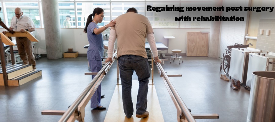
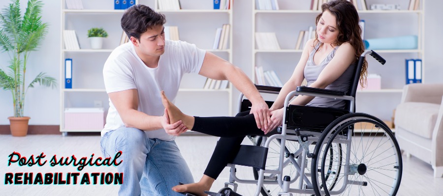
Mobility Mastery: Regaining Movement Post-Surgery with Rehabilitation

Introduction

Recovering from surgery is more than just resting; it’s a critical period where the right rehabilitation plan can make all the difference. At Arunalaya HealthCare, we understand the importance of a well-structured rehabilitation program to help patients regain strength, mobility, and confidence. Whether you’re searching for a physiotherapist for home visits in Patel Nagar, looking for advanced physiotherapy in Delhi, or need specialized care at our physiotherapy center in Delhi, our comprehensive post-surgical rehabilitation programs are designed to guide you through every phase of recovery. This blog will walk you through the essential phases of post-surgical rehabilitation, explaining how each stage contributes to your overall recovery.

Phases of Post-Surgical Rehabilitation

1. Immediate Post-Operative Phase (Days to Weeks)

Through passive range-of-motion exercises and early ambulation with assistive devices, patients can begin their journey to recovery with confidence. At Arunalaya HealthCare, our experts provide personalized care, whether at our physiotherapy center in Delhi or through physiotherapy for home visits in Inderpuri and other areas.

2. Acute Recovery Phase (Weeks 1-6)

As you transition into the acute recovery phase, the focus shifts to facilitating tissue healing, minimizing swelling, and improving joint mobility. This stage involves a gradual introduction of active range-of-motion exercises and controlled weight-bearing activities. Our specialized services, including physiotherapy in Pitampura and Punjabi Bagh, ensure that your recovery is on track with targeted interventions.

3. Intermediate Phase (Weeks 6-12)

During the intermediate phase, the goal is to increase strength, stability, and functional mobility. Progressive strength training, balance exercises, and functional activities that mimic daily tasks are introduced. Whether you’re recovering at home or at our sports rehab center in Patel Nagar, we tailor each program to your specific needs.

4. Advanced Rehabilitation Phase (Months 3-6)

The advanced phase focuses on enhancing overall strength, endurance, and specific functional abilities. This stage includes sport-specific exercises, plyometric training, and increased cardiovascular conditioning. At Arunalaya HealthCare, we provide advanced physiotherapy in Delhi, ensuring that you regain your full functional abilities with confidence.

5. Return to Activity Phase (Months 6 and Beyond)

As you approach the return to activity phase, the focus is on gradually resuming normal activities, whether it’s sports, work, or daily routines. We closely monitor your progress to prevent any signs of regression or complications, ensuring a smooth transition back to your active lifestyle.

6. Maintenance and Prevention Phase (Ongoing)

Our maintenance programs include home exercises for continued strength and flexibility, along with periodic check-ups at our physiotherapy center in Delhi. Long-term lifestyle modifications are also introduced to ensure optimal health and well-being.

Benefits of Post-Surgical Rehabilitation

Enhanced Healing and Pain Management

Post-surgical rehabilitation is crucial for reducing complications and optimizing tissue healing. Our programs focus on minimizing the need for pain medications through targeted exercises and therapeutic interventions. Whether you need chest physiotherapy near you or specialized pain management strategies, Arunalaya HealthCare is here to support you.

Restored Functionality and Prevention of Muscle Atrophy

Rehabilitation plays a key role in restoring joint flexibility, enhancing muscle strength, and preventing muscle atrophy. Our physiotherapists, available for home visits in Delhi, focus on exercises that rebuild weakened muscles, improve joint stability, and restore overall functionality.

Reduced Disability and Optimized Cardiovascular Health

Functional exercises tailored to daily life activities help reduce dependence on others, while gradual cardiovascular conditioning improves heart health and endurance. Our rehabilitation programs, available at our center and through home visits, are designed to enhance your overall quality of life.

Psychosocial Benefits and Risk Reduction

Rehabilitation also offers emotional support, helping patients maintain a positive mindset during their recovery. By addressing mobility and strength deficits, our programs reduce the risk of secondary complications and enhance joint protection, ensuring a safer, more effective recovery.

Individualized Care and Long-Term Maintenance

At Arunalaya HealthCare, we believe in personalized care. Our rehabilitation programs are tailored to each patient’s needs, ensuring that the exercises and strategies align with their specific surgical outcomes. We also emphasize long-term maintenance through continued exercise and preventive strategies to sustain the gains achieved during rehabilitation.

Conclusion

Post-surgical rehabilitation is not just about physical recovery; it’s about regaining your independence and confidence.

At Arunalaya HealthCare, we provide comprehensive rehabilitation programs that cater to each phase of your recovery, whether you’re at home or visiting our physiotherapy center in Delhi. With services like physiotherapy for home visits in Patel Nagar, advanced physiotherapy in Delhi, and specialized care in areas like Pitampura and Punjabi Bagh, we are committed to helping you achieve the best possible outcomes. Embrace your journey to recovery with Arunalaya HealthCare, and let us guide you towards a healthier, more active life.


physiotherapy for home visit in patel nagar | physiotherapy in pitampura | best physiotherapist in rajender nagar | physiotherapy in naraina | best physiotherapist in inderpuri | physiotherapy center in punjabibagh | physiotherapy in punjabi bagh| physiotherapy in karol bagh | sports injury centre in delhi | sports physiotherapist in patel nagar | sports rehab center in patel nagar | cp treatment | physio clinic near me in delhi | chest physiotherapy near me | advanced physiotherapy in delhi | best sports injury physiotherapist in delhi | best sports injury physiotherapy in delhi | sports physiotherapist in delhi | back pain physiotherapy near me | home visit physiotherapy in delhi | physiotherapy center in delhi | best physiotherapist at home in patel nagar | physiotherapy in patel nagar | physiotherapist in patel nagar

best sports injury physiotherapist in delhi

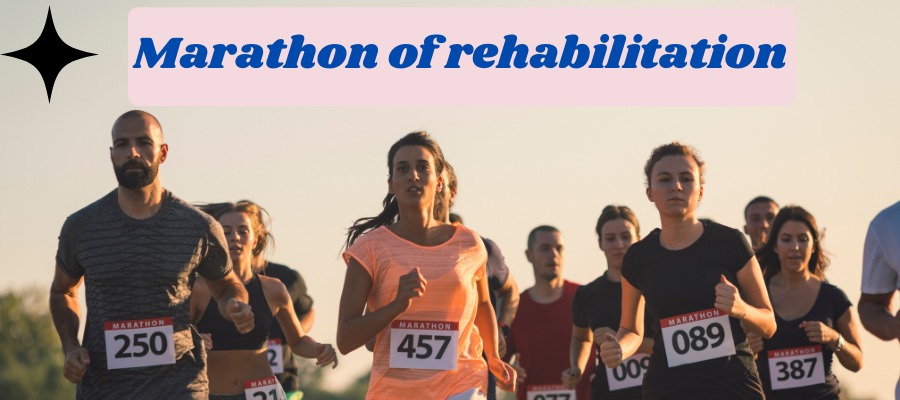
The Marathon of Rehabilitation: Endurance Strategies in Sports Physiotherapy

Introduction

Recovering from a marathon is not just about resting; it’s a structured process that requires thoughtful endurance strategies. At Arunalaya HealthCare, we understand that athletes need more than just time off after such an intense event. Our approach incorporates advanced physiotherapy techniques, ensuring a comprehensive recovery that prepares the body for the next challenge. Whether you need physiotherapy for home visits in Patel Nagar or are searching for a physiotherapy center in Delhi, we provide personalized care tailored to your unique needs. This blog explores the stages of post-marathon recovery and the endurance strategies that can help you return to peak performance.

Immediate Post-Marathon Care

The first moments after crossing the marathon finish line are crucial. At Arunalaya HealthCare, we emphasize the importance of keeping your body moving for 10-15 minutes post-race. This helps in maintaining blood flow and reducing the risk of muscle stiffness. Avoid static stretching, as the muscle damage you experience can worsen with premature stretching. Instead, focus on gentle, dynamic movements to ease your body back to normalcy.

The First Hour After the Race

Within an hour, it’s vital to replenish your energy stores with a carb-rich snack or drink. At our physiotherapy center in Delhi, we often recommend that marathon runners change out of their damp clothing as soon as possible to avoid lingering discomfort and further stress on the body.

The Afternoon and Evening After the Marathon

Sleep is a key component of muscle repair, and a short nap of up to 90 minutes can significantly aid recovery. Avoid anti-inflammatory painkillers like ibuprofen, as they can cause liver damage post-race. Instead, opt for safer alternatives like paracetamol if you’re in pain. Before bed, a milk-based drink can provide the carbohydrates and proteins necessary for overnight recovery. Remember, while a celebratory beer is fine, excessive alcohol can slow down your recovery.

The Day After the Marathon

Compression socks or tights can be a runner’s best friend the day after a marathon. They help boost recovery by improving circulation. It’s also important to protect your immune system, which is weakened post-race. Avoid close contact with anyone who has a cold or infection to prevent catching something while your body is vulnerable.

Two Days After the Marathon

Active recovery, like a gentle swim, is highly beneficial two days post-marathon. Research published in the International Journal of Sports Medicine suggests that swimming-based recovery can enhance overall exercise performance. However, resist the urge to start running just yet; your body still needs time to heal.

Three to Four Days After the Marathon

A sports massage can be incredibly beneficial during this phase, but timing is crucial. At Arunalaya HealthCare, we advise against immediate post-race massages, as they can exacerbate muscle damage. Instead, opt for a massage a few days later, when your body has had some time to heal. Additionally, avoid ice baths days after the race. Warm baths are better as they promote blood flow and aid in muscle recovery.

Five to Six Days After the Marathon

Now is the time for a test run of 20 – 30 minutes. Pay close attention to any lingering aches or pains. It’s important to avoid speed work or hills during this phase. The goal is to ease back into your routine, not to push your body to the limit again too soon.

Endurance Strategies in Sports Physiotherapy

Endurance in sports is not just about physical stamina; it’s about the mental toughness and physiological adaptations that occur with consistent training. At Arunalaya HealthCare, our advanced physiotherapy in Delhi focuses on both general and sport-specific endurance. Whether you’re training for a marathon or another endurance sport, our programs are designed to improve your body’s efficiency, from aerobic capacity to neuromuscular adaptations.

Endurance training leads to significant adaptations in both skeletal muscle and the cardiovascular system. These changes enhance your body’s ability to transport and use oxygen, delay muscle fatigue, and ultimately improve performance. Through tailored endurance strategies, including advanced physiotherapy techniques, we help athletes of all levels achieve their peak performance.

Conclusion

Recovery after a marathon is a journey that requires endurance, not just during the race, but long after. At Arunalaya HealthCare, we support this journey with comprehensive physiotherapy services that cater to every aspect of your recovery. Whether you’re looking for physiotherapy for home visits in Patel Nagar, or a sports rehab center in Patel Nagar, our team is here to guide you every step of the way. By following the right recovery strategies and endurance training, you can return to your regular running routine stronger and more resilient than ever before.

“At Arunalaya Healthcare, we pride ourselves on being the best physiotherapy center in Delhi. Our dedicated team of experts offers top-notch physiotherapy treatment tailored to your needs. Experience the difference with the leading physiotherapy clinic in Delhi area. Our commitment to excellence ensures that you receive the best physiotherapy care possible. Trust Arunalaya Healthcare for the best physiotherapy treatment in Delhi. Our advanced physiotherapy solutions set us apart as the premier choice for your rehabilitation needs. When it comes to physiotherapy, our center stands out as the best in Delhi. Choose Arunalaya Healthcare for comprehensive physiotherapy solutions that deliver results. Visit Arunalaya Healthcare today and discover why we are the best physiotherapy center in Delhi.”

physiotherapy for home visit in patel nagar | physiotherapy in pitampura | best physiotherapist in rajender nagar | physiotherapy in naraina | best physiotherapist in inderpuri | physiotherapy center in punjabibagh | physiotherapy in punjabi bagh| physiotherapy in karol bagh | sports injury centre in delhi | sports physiotherapist in patel nagar | sports rehab center in patel nagar | cp treatment | physio clinic near me in delhi | chest physiotherapy near me | advanced physiotherapy in delhi | best sports injury physiotherapist in delhi | best sports injury physiotherapy in delhi | sports physiotherapist in delhi | back pain physiotherapy near me | home visit physiotherapy in delhi | physiotherapy center in delhi | best physiotherapist at home in patel nagar | physiotherapy in patel nagar | physiotherapist in patel nagar

physiotherapy in delhi

Path to Recovery: Effective Post-Surgical Rehabilitation

Introduction

Post-surgical rehabilitation combines the art and science of therapeutic techniques to optimize recovery and functionality following surgery. At Arunalaya HealthCare, we understand that every individual’s needs and goals are unique, and we tailor our programs to address these differences. Whether you require physiotherapy for home visits in Patel Nagar or advanced physiotherapy in Delhi, our team is dedicated to supporting your journey to wellness.

Assessment and Evaluation

This includes:

Comprehensive Evaluation: Reviewing the patient’s medical history, including previous surgeries, medical conditions, medications, and allergies.

Detailed Surgical Assessment: Understanding the type of surgery performed, surgical approach, and any intraoperative complications.

Physical Examination: Assessing range of motion, strength, joint stability, sensation, and overall function.

Outcome Measures: Utilizing functional assessments to establish a baseline and track progress.

Psychosocial Consideration: Factoring in patient expectations, motivation, social support, and potential barriers to recovery.

Pain Management

Effective pain management is a cornerstone of post-surgical rehabilitation.

Individualized Plans: Tailored pain management strategies based on pain type and severity, patient preferences, and potential medication side effects.

Multimodal Analgesia: Combining pharmacological and non-pharmacological interventions to maximize pain relief and minimize adverse effects.

Inflammation Reduction: Using ice, heat, compression, and elevation techniques to alleviate acute postoperative pain.

Chronic Pain Strategies: Implementing relaxation techniques, biofeedback, mindfulness-based interventions, and cognitive-behavioral strategies for persistent pain.

Range of Motion (ROM) Exercises

Restoring flexibility and joint mobility is essential.

Gradual Progression: Moving from passive ROM exercises to active-assisted and active exercises as tolerated.

Stretching and Mobilization: Using stretching techniques, joint mobilizations, and proprioceptive neuromuscular facilitation (PNF).

CPM Devices: Employing continuous passive motion devices and manual therapy to prevent adhesions and maintain joint integrity.

Strength Training

Building strength post-surgery involves.

Targeted Programs: Focused on muscle groups affected by surgery to restore functional strength, endurance, and power.

Progressive resistance: training using free weights, resistance bands, machines, and bodyweight exercises to stimulate muscle hypertrophy and neuromuscular adaptation.

Balanced Contractions: Integrating eccentric, concentric, and isometric contractions to address muscle imbalances and weaknesses.

Functional Training

Enhancing daily activities and sports performance.

Task-Specific Exercises: Improving functional capacity, motor control, and movement patterns.

Balance and Agility: Incorporating balance challenges, coordination drills, and dynamic movements.

Real-Life Simulations: Facilitating transitions back to home, work, and recreational activities.

Manual Therapy

Promoting tissue healing and flexibility.

Tailored Techniques: Based on tissue healing stages, pain levels, and mobility restrictions.

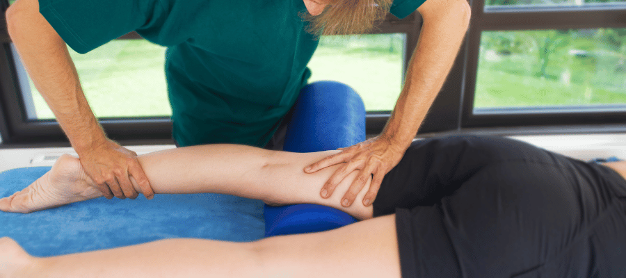
Soft Tissue Mobilization: Including massage, psychological release, scar massage, and trigger point therapy.

Joint Mobilizations: Restoring normal joint movements and alleviating stiffness.

Balance and Proprioception Training

Improving stability and preventing falls.

Progressive Exercises: On stable and unstable surfaces to challenge sensory systems.

Sensory Integration: Enhancing postural stability and dynamic equilibrium.

Perturbation Training: Improving reaction time and fall prevention strategies.

Education and Counseling

Empowering patients with knowledge and support.

Information Provision: About the surgical procedure, recovery timeline, potential complications, and precautions.

Ergonomic Guidance: Teaching proper body mechanics and activity modification.

Psychological Support: Addressing fears, anxieties, and emotional distress related to surgery and rehabilitation.

Progressive Overload

Gradual increase in exercise demands.

Systematic Progression: Of exercise intensity, volume, and complexity to promote tissue remodeling and performance gains.

Rehabilitation Phases: Acute, subacute, and chronic phases with adjustments based on individual responses.

Regular Monitoring: To track progress, identify barriers, and modify treatment strategies.

Monitoring and Adjustment

Ongoing assessment to ensure optimal recovery.

Patient Response: Monitoring pain levels, functional outcomes, adherence to home exercises, and activity compliance.

Reassessment: Regularly evaluating joint mobility, muscle strength, and functional capacity to adjust goals as needed.

Conclusion

Embarking on the path to recovery after surgery can be a challenging journey, but with the right approach and support, optimal healing and functionality are within reach. At Arunalaya HealthCare, we prioritize personalized rehabilitation plans that encompass comprehensive assessments, pain management strategies, and progressive strength training. Our holistic approach integrates functional training, manual therapy, and balance exercises, ensuring a well-rounded recovery. With continuous monitoring and adjustments, we aim to empower our patients with the knowledge and tools needed for sustained recovery and a return to their daily activities.

Post-surgical rehabilitation at Arunalaya HealthCare is designed to help patients recover effectively and safely. Whether you need physiotherapy for home visits in Patel Nagar, advanced physiotherapy in Delhi, or specialized treatments like CP or chest physiotherapy, our dedicated team is here to support you every step of the way.


physiotherapy for home visit in patel nagar | physiotherapy in pitampura | best physiotherapist in rajender nagar | physiotherapy in naraina | best physiotherapist in inderpuri | physiotherapy center in punjabibagh | physiotherapy in punjabi bagh| physiotherapy in karol bagh | sports injury centre in delhi | sports physiotherapist in patel nagar | sports rehab center in patel nagar | cp treatment | physio clinic near me in delhi | chest physiotherapy near me | advanced physiotherapy in delhi | best sports injury physiotherapist in delhi | best sports injury physiotherapy in delhi | sports physiotherapist in delhi | back pain physiotherapy near me | home visit physiotherapy in delhi | physiotherapy center in delhi | best physiotherapist at home in patel nagar | physiotherapy in patel nagar | physiotherapist in patel nagar

Floating Knee

Floating Knee

Table Of Contents

  • Introduction
  • What is a Floating Knee?
  • Types of Floating Knee Injuries
  • Complications of Floating Knee Injuries
  • Management of Floating Knee Injuries
  • Conclusion

Introduction

A floating knee is a severe injury involving fractures of both the femur (thigh bone) and tibia (shin bone) in the same leg, creating a “floating” joint. This condition, usually caused by high-energy trauma such as car accidents or falls from significant heights, was first described by Blake and McBryde in 1975. It represents a rare and complex injury requiring prompt and specialized treatment to prevent severe complications.

What is a Floating Knee?

A floating knee results from simultaneous fractures of the femur and tibia on the same leg, leading to a flail knee joint characterized by instability due to damaged supporting ligaments. This instability can cause pain, swelling, and difficulty in movement. The high incidence of open fractures, vascular injuries, and nerve damage makes this condition particularly challenging to manage.

Types of Floating Knee Injuries

Floating knee injuries have been classified using various systems to better understand and treat the condition. Here are some key classification systems:

Blake and McBryde Classification

  1. Type I: Fractures of both shafts of the two long bones.
  2. Type II-A: Fractures involving the knee joint.
  3. Type II-B: Fractures involving the hip or ankle joints.

Letts-Vincent Classification (Pediatric)

  1. Type A: Two closed diaphyseal fractures.
  2. Type B: One closed diaphyseal and one metaphyseal fracture.
  3. Type C: One diaphyseal and one epiphyseal fracture.
  4. Type D: At least one open fracture.
  5. Type E: Both fractures are open.

Bohn-Durbin Classification (Pediatric)

  1. Type I: Double shaft fractures.
  2. Type II: Juxta-articular injuries.
  3. Type III: Fractures with an epiphyseal component.

Fraser Classification

  1. Type I: Shaft fractures of both bones without knee involvement.
  2. Type II: Fractures extending into the knee.
    • Type II a: Tibial plateau involvement.
    • Type IIb: Distal femur involvement.
    • Type II c: Both tibial plateau and distal femur involvement within the knee joint.

Complications of Floating Knee Injuries

Floating knee injuries can lead to various complications, including:

  • Epiphyseal injury: May affect open growth plates in children, causing limb-length discrepancies and deformities.
  • Infections: Due to open fractures.
  • Nonunion and malunion: Improper healing of fractures.
  • Knee stiffness: Resulting in functional impairment.

Management of Floating Knee Injuries

Immediate medical attention and stabilization are critical for patients with floating knees to prevent further damage and address associated injuries. Treatment typically involves surgical stabilization of the fractures, followed by a comprehensive rehabilitation program.

Physiotherapy Rehabilitation

  • Post-surgical Examination: Assess range of motion and stability.
  • Ligament Reconstruction: Usually delayed until skeletal injury rehabilitation is adequate.
  • Pain Control: Essential in the early postoperative phase, often managed with epidural catheters or systemic opioid infusion.
  • Weight Bearing:
    • Type I: Delayed until callus formation is visible on radiographs.
    • Type II: Permitted only after 10 weeks.
  • Early Mobilization: Crucial for optimal recovery, especially in cases involving intra-articular fractures.

Conclusion

Floating knee injuries represent a complex and severe form of trauma requiring prompt, specialized medical intervention and comprehensive rehabilitation. Understanding the types, complications, and management strategies for floating knees is essential for healthcare providers to ensure the best possible outcomes for patients. With proper treatment and rehabilitation, patients can regain function and mobility, minimizing long-term complications.


“At Arunalaya Healthcare, we pride ourselves on being the best physiotherapy center in Delhi. Our dedicated team of experts offers top-notch physiotherapy treatment tailored to your needs. Experience the difference with the leading physiotherapy clinic in Delhi area. Our commitment to excellence ensures that you receive the best physiotherapy care possible. Trust Arunalaya Healthcare for the best physiotherapy treatment in Delhi. Our advanced physiotherapy solutions set us apart as the premier choice for your rehabilitation needs. When it comes to physiotherapy, our center stands out as the best in Delhi. Choose Arunalaya Healthcare for comprehensive physiotherapy solutions that deliver results. Visit Arunalaya Healthcare today and discover why we are the best physiotherapy center in Delhi.”


Physiotherapist in Patel Nagar | Physiotherapist for Home Visit in Patel Nagar | Physiotherapy in Patel Nagar | Best Physiotherapist in Patel Nagar | Physiotherapist Near Me | Physiotherapy Near Me | Best Physiotherapist in Delhi | Best Physiotherapist in India | Physiotherapy Center in Patel Nagar | Spine Clinic Near Me | Back Pain Physiotherapy Near Me | Sports Physiotherapist in Delhi | Stroke Physiotherapy | Paralysis | Cerebral Palsy | Best Sports Injury Physiotherapist in Delhi | Best Sports Injury Physiotherapy in Delhi | Physiotherapy Home Service | Physiotherapy at Home | Home Visit Physiotherapy | Advanced Physiotherapy in Delhi | Physiotherapy Clinic Near Me | Chest Physiotherapy Near Me

How to Restore Knee Extension Range of Motion After ACL Reconstruction.

Table Of Contents

  • Introduction
  • Some of The Most Important and Simple Exercises to Improve Knee Extension Range Following ACL Reconstruction
  • Tips to Consider
  • Conclusion

Introduction

Knee extension deficit or loss of extension is a potential complication following ACL reconstruction. Lack of full extension after an ACL rupture may contribute to functional deficits and an increased risk of osteoarthritis. Loss of terminal extension often occurs because of hamstring contracture and quadriceps inactivation. Failure to regain full extension during the first few weeks may pose a risk for adverse long-term outcomes, making it crucial to address the problem at the earliest.

Some of The Most Important and Simple Exercises to Improve Knee Extension Range Following ACL Reconstruction

  1. Heel Raise
    • One of the best, most passive ways to work on extension is by sitting with your leg and raising your heel.
  2. Quads Squeeze with Heel Raise
    • Sit straight and try to lay your leg flat and wrap a towel around your heel. As you squeeze your quad, try to pull the towel up to help straighten your leg. Hold for 30-60 seconds and relax.
  3. Elevated Leg Extensions on Chair
    • Sit on one chair across from another chair of the same height. Straighten your leg so that your heel is on the heel across from you, and make sure there is no chair under your knee.
  4. Prone Hang
    • Lay on your stomach on a bed or table, with the end of the surface hitting the quad just above the knee. Relax and let your leg straighten.
  5. Walking Straight
    • Your walking is really important for ROM. As you walk, put your leg all the way straight. Try walking backwards as well to straighten the leg with motion.
  6. Terminal Knee Extension with Band
    • This is a great exercise for knee extension. Attach a band to something stationary, step into the band, placing it on the hamstring just above your knee. Push your knee back against the band to straighten your leg as much as possible.
  7. Squats with Calf Raises
    • While squatting, try to come up on your toes and go into a calf raise while pushing your leg as much as possible.
  8. Hamstring Stretch
    • Do at least 3-4 times, holding for 30-60 seconds.
  9. Slant Board
    • Use a slant board to elongate your calves and push your knee as straight as possible. Hold for 30-60 seconds and as you improve extension, select a more and more steep slant board.

Tips to Consider

  1. You must complete the exercises for knee extension several times a day.
  2. Listen to your body.
  3. Don’t overstretch.
  4. Don’t neglect flexion. Work on both types of ROM.

Conclusion

Restoring knee extension range of motion after ACL reconstruction is crucial to prevent functional deficits and long-term complications like osteoarthritis. By incorporating simple exercises such as heel raises, quad squeezes, elevated leg extensions, prone hangs, walking straight, terminal knee extensions with bands, squats with calf raises, hamstring stretches, and using a slant board, patients can improve their knee extension. It’s essential to follow these exercises diligently, listen to your body, avoid overstretching, and not neglect flexion exercises to achieve the best outcomes in restoring knee function.


“At Arunalaya Healthcare, we pride ourselves on being the best physiotherapy center in Delhi. Our dedicated team of experts offers top-notch physiotherapy treatment tailored to your needs. Experience the difference with the leading physiotherapy clinic in Delhi area. Our commitment to excellence ensures that you receive the best physiotherapy care possible. Trust Arunalaya Healthcare for the best physiotherapy treatment in Delhi. Our advanced physiotherapy solutions set us apart as the premier choice for your rehabilitation needs. When it comes to physiotherapy, our center stands out as the best in Delhi. Choose Arunalaya Healthcare for comprehensive physiotherapy solutions that deliver results. Visit Arunalaya Healthcare today and discover why we are the best physiotherapy center in Delhi.”


Physiotherapist in Patel Nagar | Physiotherapist for Home Visit in Patel Nagar | Physiotherapy in Patel Nagar | Best Physiotherapist in Patel Nagar | Physiotherapist Near Me | Physiotherapy Near Me | Best Physiotherapist in Delhi | Best Physiotherapist in India | Physiotherapy Center in Patel Nagar | Spine Clinic Near Me | Back Pain Physiotherapy Near Me | Sports Physiotherapist in Delhi | Stroke Physiotherapy | Paralysis | Cerebral Palsy | Best Sports Injury Physiotherapist in Delhi | Best Sports Injury Physiotherapy in Delhi | Physiotherapy Home Service | Physiotherapy at Home | Home Visit Physiotherapy | Advanced Physiotherapy in Delhi | Physiotherapy Clinic Near Me | Chest Physiotherapy Near Me

Understanding Arthrofibrosis and its Management through Physiotherapy

Table Of Contents

  • Introduction
  • Understanding Arthrofibrosis
  • Causes of Arthrofibrosis
  • Physiotherapy’s Role in Arthrofibrosis Management
  • Conclusion

Introduction

Arthrofibrosis is a medical condition characterized by the excessive formation of scar tissue within a joint, leading to a loss of motion and function. This condition can be debilitating, affecting individuals who have undergone joint surgery, experienced trauma, or suffered from chronic inflammation. In this blog, we will delve into what arthrofibrosis is, its causes, and how physiotherapy plays a crucial role in its treatment and management.

Understanding Arthrofibrosis

Arthrofibrosis primarily affects the knee, although it can occur in other joints like the shoulder, elbow, or hip. The condition develops when the body’s natural response to injury, surgery, or inflammation leads to an overproduction of scar tissue within the joint capsule. This excess scar tissue can restrict joint movement, causing stiffness, pain, and a decrease in overall joint function.

Causes of Arthrofibrosis

Several factors can contribute to the development of arthrofibrosis:

  1. Surgery: Post-operative scarring and inflammation can lead to arthrofibrosis, particularly in procedures involving joint surgeries such as knee replacements or ACL reconstructions.
  2. Trauma: Injuries like fractures or dislocations can trigger excessive scar tissue formation during the healing process.
  3. Chronic Inflammation: Conditions like rheumatoid arthritis can lead to ongoing joint inflammation, increasing the risk of arthrofibrosis.
  4. Infection: Joint infections can cause inflammation and scarring within the joint.
  5. Poor Rehabilitation: Inadequate or improper post-injury or post-surgery rehabilitation can contribute to arthrofibrosis.

Physiotherapy’s Role in Arthrofibrosis Management

Physiotherapy is a cornerstone in the management and treatment of arthrofibrosis. A well-designed physiotherapy plan can help alleviate symptoms, improve joint mobility, and prevent the recurrence of scar tissue formation. Here’s how physiotherapy can aid in arthrofibrosis management:

  1. Early Intervention: Timely physiotherapy can begin shortly after surgery or injury, aiming to prevent the excessive buildup of scar tissue by promoting proper joint mobilization and range of motion exercises.
  2. Range of Motion Exercises: Physiotherapists employ specific exercises to improve joint flexibility and reduce stiffness. Passive, active-assisted, and active range of motion exercises are tailored to the individual’s condition.
  3. Strengthening Exercises: Strengthening the muscles around the affected joint can enhance stability and support, reducing the strain on the joint itself. This can also prevent the development of compensatory movement patterns.
  4. Manual Therapy: Hands-on techniques, such as joint mobilization and soft tissue manipulation, can help break down scar tissue, improve circulation, and restore joint function.
  5. Pain Management: Physiotherapists utilize various modalities like ultrasound, heat, or ice to manage pain and inflammation associated with arthrofibrosis.
  6. Education and Home Exercises: Patients are educated on self-management techniques and provided with home exercise programs to continue their rehabilitation outside of clinic visits.
  7. Gradual Progression: Physiotherapy plans are tailored to the patient’s progress, ensuring that exercises and interventions are gradually intensified to prevent re-accumulation of scar tissue.
  8. Postural Correction: Correcting posture and movement patterns is crucial to reduce joint stress and prevent the development of arthrofibrosis.

Conclusion

Arthrofibrosis is a challenging condition that can significantly impact an individual’s quality of life. However, with a well-structured physiotherapy plan, patients can experience significant improvements in joint mobility and function. Early intervention, proper exercise protocols, and ongoing support from physiotherapists are essential elements in managing arthrofibrosis effectively. If you or someone you know is dealing with arthrofibrosis, consult a qualified physiotherapist to develop a personalized treatment plan tailored to your specific needs.


“At Arunalaya Healthcare, we pride ourselves on being the best physiotherapy center in Delhi. Our dedicated team of experts offers top-notch physiotherapy treatment tailored to your needs. Experience the difference with the leading physiotherapy clinic in Delhi area. Our commitment to excellence ensures that you receive the best physiotherapy care possible. Trust Arunalaya Healthcare for the best physiotherapy treatment in Delhi. Our advanced physiotherapy solutions set us apart as the premier choice for your rehabilitation needs. When it comes to physiotherapy, our center stands out as the best in Delhi. Choose Arunalaya Healthcare for comprehensive physiotherapy solutions that deliver results. Visit Arunalaya Healthcare today and discover why we are the best physiotherapy center in Delhi.”


Physiotherapist in Patel Nagar | Physiotherapist for Home Visit in Patel Nagar | Physiotherapy in Patel Nagar | Best Physiotherapist in Patel Nagar | Physiotherapist Near Me | Physiotherapy Near Me | Best Physiotherapist in Delhi | Best Physiotherapist in India | Physiotherapy Center in Patel Nagar | Spine Clinic Near Me | Back Pain Physiotherapy Near Me | Sports Physiotherapist in Delhi | Stroke Physiotherapy | Paralysis | Cerebral Palsy | Best Sports Injury Physiotherapist in Delhi | Best Sports Injury Physiotherapy in Delhi | Physiotherapy Home Service | Physiotherapy at Home | Home Visit Physiotherapy | Advanced Physiotherapy in Delhi | Physiotherapy Clinic Near Me | Chest Physiotherapy Near Me