Shock Wave Therapy: How it Works?

Shock Wave therapy is a technique that uses high-energy sound waves rather than electrical shocks to aid in the healing process following an injury.

SWT is a cutting-edge treatment that uses acoustic shock waves to break up soft tissue calcifications, boost collagen production, release growth factors, and accelerate your body’s healing process in order to reduce pain and keep you active.

It’s a lesser-known physiotherapy technique, yet it can be beneficial.

Injuries that can be treated

Shock wave therapy is used to treat musculoskeletal injuries, particularly those areas involving connective tissue and bone.
The following are some examples of common injuries treated with shockwave therapy:

  • tennis/golf elbow
  • rotator cuff tendonitis
  • calcification
  • stress fractures
  • trochanteric bursitis in the hip
  • patellar tendonitis
  • jumper’s knee
  • hell spurs
  • Achilles tendonitis
  • Morton’s neuroma in the foot

Shock wave therapy treatment has been shown to be an excellent approach to start the body’s healing process and reduce discomfort while regaining movement.
Most people require only three treatments per week.

This therapy improves blood circulation and hastens the repair of chronic musculoskeletal disorders in the bones, tendons, ligaments, and muscle.

How does it work?

  • Shockwave treatment stimulates self-healing by using radial acoustic shockwaves.
  • These shockwaves, which are not electrical but rather sound shockwaves, are administered through a special wand hand piece that dispenses the waves for a brief amount of time directly to the affected injured tissue areas.
  • The pressure from the shockwaves will be transferred to the tissue. As a result, special micro-cavitation bubbles form, expand, burst, and produce a force known as a resultant force.
  •  This passes through the tissue and stimulates the cells responsible for connective tissue and bone mending, so activating the body’s inherent self-healing mechanism.
  • These processes involve increased metabolism and blood circulation. It means that your damaged tissue receives better healing treatment from your body, and the damaged tissue will begin to regenerate itself, with an accompanying inflammatory response allowing the healing process to take effect.
  • Some people find it slightly uncomfortable to go through. However, these people are frequently in the minority, and the majority are able to handle the five-minute treatments with ease.
  • Though there will be no pain immediately following the treatment, there is a potential of discomfort in the hours that follow.
  • This discomfort can last for up to two days, therefore patients should limit their physical activity and avoid using anti-inflammatory drugs or ice.

How does it feel?

ESWT hurts, and the degree of pain varies from person to person. The majority of patients perceive it as slightly uncomfortable but not painful.
It also affects where we treat you. Body portions with less skin and muscle, such as above the hip bone, can be more sensitive than meatier areas, such as the calves.

OSTEOPOROSIS MANAGEMENT IN PHYSIOTHERAPY

Osteoporosis is a condition in which the bones in the body become weak and brittle as a result of tissue loss over time. This illness is most common in the elderly, but it can also occur in middle-aged patients.

Although this condition can affect any bone in the body, it most commonly affects the following:

  • Spine
  • Hips
  • Waist

Because of bone fragility, people with osteoporosis are susceptible to fractures.

Causes

While weak bone structures are caused by ageing factors, smoking, and excessive alcohol use, a prolonged sedentary lifestyle can also play a role. Cigarettes contain toxic substances that interfere with the bones’ natural ability to repair, while alcohol interferes with calcium absorption.

Habitual physical inactivity promotes the activity of osteoclasts (the cells responsible for bone resorption).

Another factor that contributes to osteoporosis is hormonal change associated with ageing. Women going through menopause may be more likely to develop osteoporosis later in life due to lower oestrogen levels (a hormone that helps manufacture bone mass).

Anyone can get osteoporosis, and the risk rises with age.

Role of Physiotherapy

Physiotherapy can aid in the prevention and management of osteoporosis.
Over the course of weeks and months, a physical therapist will design a personalised training programme to strengthen your bones and muscles.
This improves your balance and reduces your chances of falling.

Physical therapy can also aid in the recovery from an osteoporosis-related accident.

Physical therapy often involves performing stretches or exercises, usually done in repetition or sets. But physical therapists also use a variety of other techniques to improve movement.

These include:

  • Manual therapy
  • Kinesiology taping
  • Dry needling
  • suggestions for lifestyle changes
  • MFR
  • heat or cold therapy
  • ultrasound / electrical stimulation
  • Stability training

Benefits of PT

  • Physiotherapy treatments aid in the strengthening of bones and muscles
  • Regular and targeted exercise is essential for the prevention and management of osteoporosis.
  • Working with a physiotherapist can help you with the following:
    1. improve your balance
    2. lower your chances of falling
    3. stretch and strengthen your muscles
    4. correct stooped posture
    5. prevent further bone loss
    6. reduce pain

Understanding Lateral Ligament Injury of the Ankle

Lateral ligament injury is among the most prevalent types of sports injuries addressed by physiotherapists. Men and women are estimated to suffer from lateral ankle sprains at roughly the same rates.

A lateral ligament injury of the ankle is a common injury that occurs when the ligaments on the outer side of the ankle are damaged. The most frequently affected ligaments are the anterior talofibular ligament (ATFL), the calcaneofibular ligament (CFL), and the posterior talofibular ligament (PTFL). These ligaments are responsible for providing stability to the ankle joint.

Lateral ankle sprains are also known as inversion or supination ankle sprains. The complex of ligaments on the lateral side of the ankle is torn by varied degrees as a result of a forced plantarflexion/inversion movement.

Individuals who sustain multiple recurrent ankle sprains have been documented to have functional and mechanical instability, as well as an increased risk of re-injury. Small fractures surrounding the ankle and foot (e.g. Pott’s fracture) and straining or rupture of the muscles around the ankle (e.g. calf, peroneii, tibialis anterior) should also be avoided even if they are the less prevalent causes of ankle pain.

Lateral Ankle Ligament Tear Types:

Lateral ligament injuries of the ankle can be classified into three main types based on the ligaments involved and the severity of the tear:

Anterior talofibular ligament (ATFL) injury: This is the most common type of lateral ligament injury and often occurs in isolation. It involves the stretching or tearing of the ATFL, which connects the talus bone to the fibula. It is commonly associated with ankle inversion injuries.

Calcaneofibular ligament (CFL) injury: The CFL is located slightly below the ATFL and connects the calcaneus (heel bone) to the fibula. CFL injuries typically occur along with ATFL injuries or in more severe sprains. In some cases, the ATFL and CFL can be injured together, resulting in a higher-grade ligament tear.

Posterior talofibular ligament (PTFL) injury: The PTFL is the least commonly injured ligament in lateral ligament injuries of the ankle. It connects the talus bone to the fibula at the back of the ankle. PTFL injuries usually occur in severe ankle sprains or high-energy trauma.

CAUSES

Ankle sprain: The most common cause of a lateral ligament injury is an ankle sprain, which typically occurs when the foot rolls inward, resulting in stretching or tearing of the ligaments on the outer side of the ankle.

Sports injuries: Activities that involve quick changes in direction, jumping, or running on uneven surfaces increase the risk of ankle sprains and ligament injuries.

Trauma: Direct trauma or impact to the ankle joint, such as a fall or a collision, can cause damage to the lateral ligaments.

CLINICAL FEATURES

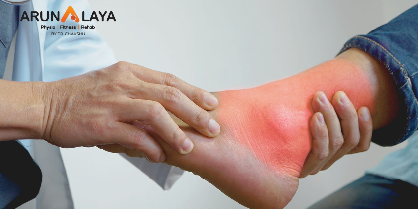
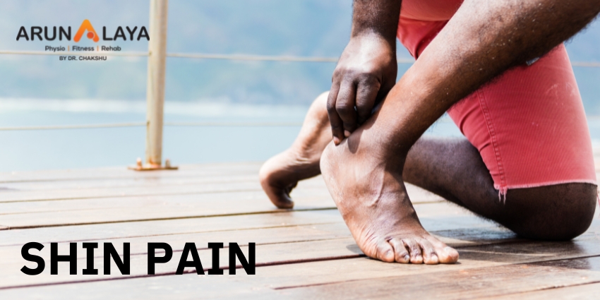
  • Pain on the outer side of the ankle
  • Swelling and bruising
  • Difficulty walking or bearing weight on the affected foot
  • Instability or a feeling of the ankle giving way
  • Limited range of motion in the ankle joint.

RISK FACTORS

Body mass index, slow eccentric inversion strength, quick concentric plantar flexion strength, passive inversion joint position sense, and peroneus brevis reaction time were all linked to an elevated risk of lateral ankle injury.

ASSESSMENT OF ANKLE JOINT

  • Amount of instability present by assessing the grade of the sprain;
  • Loss of Range of motion (ROM);
  • Loss of the muscle strength;
  • Level of reduced Proprioception.

OBSERVATION

Any symptoms of injury, inflammation, skin colour changes, or muscle atrophy or hypertrophy are noted. Following that, the foot and ankle are observed in two separate positions: non-weight bearing and weight bearing. Take note of the gait pattern, degree of limp (if any), and facial expression when bearing weight.

HISTORY

Mechanism of injury: A plantarflexion/inversion injury would indicate damage to the lateral ligament, whereas a dorsiflexion/eversion injury would indicate damage to the medial ligament. Previous history of injury on the same side will give clues as to whether the ankle was unstable to begin with, or that a previous injury wasn’t properly rehabilitated.

History of injury on the other side as well may indicate a biomechanical predisposition towards ankle injuries.

GRADES

Grade 1: Mild sprain with minimal stretching or tearing of the ligament fibers. There may be slight swelling, tenderness, and minimal loss of function.

Grade 2: Moderate sprain with partial tearing of the ligament fibers. This grade is characterized by increased swelling, bruising, pain, and difficulty walking or bearing weight on the affected ankle.

Grade 3: Severe sprain with complete tearing or rupture of the ligament. Significant swelling, bruising, pain, and instability are commonly observed. Walking or weight-bearing may be extremely difficult or impossible.

SPECIAL TEST

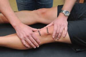
An anterior draw is performed to assess the ATFL and CFL integrity. The heel is grabbed with the ankle in plantarflexion, and the tibia is stabilised and dragged anteriorly.
The talar tilt is used to evaluate the integrity of the ATFL and CFL laterally, as well as the deltoid ligament medially. The heel is grabbed again, the tibia is stabilised, and the talus and calcaneus are pushed laterally and medially.
Beginning with a simple single leg stance, proprioception can be measured in a variety of more challenging methods. The patient can do it on the normal side first to give the therapist a sense of what is typical, and then try it on the injured side.

DIFFERENTIAL DIAGNOSIS

  • Ankle fracture (medial/lateral malleolus, distal tibia/fibular)
  • Damage to the medial ligament
  • Dislocated ankle
  • Other soft tissue damage (peroneal tendons, muscle strain)

TREATMENT

REDUCE PAIN AND SWELLING

The RICE regimen (Rest, Ice, Compression, and Elevation) can be used to minimise pain and swelling in the first 48-72 hours after an acute lateral ligament damage.

If weight bearing is too painful, the patient can use elbow crutches for 24 hours and remain non-weight bearing. However, it is critical to begin at least partial weight bearing as soon as possible, along with a regular heel-toe gait pattern, since this will help to reduce pain and swelling.

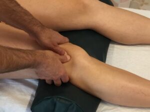
Gentle soft tissue massage and light stretches can be conducted to help with the clearance of oedema, as long as they are painless.

Range of motion exercises: These exercises aim to restore the normal range of motion in the ankle joint and may involve gentle ankle rotations, ankle pumps, and alphabet exercises.

Strengthening exercises: Strengthening the muscles around the ankle joint helps provide support and stability. Common exercises include calf raises, toe raises, ankle inversions and eversions (using resistance bands or manual resistance), and single-leg balance exercises.

Proprioceptive and balance exercises: These exercises improve the body’s awareness of joint position and enhance balance and stability. Examples include standing on one leg, balance board exercises, and wobble board exercises.

RETURN TO FUNCTIONAL ACTIVITY

  • Twisting
  • Jumping
  • Hopping on one leg
  • Running
  • Figure of 8 running

Before returning to full functional activity the patient should have full range of pain free movement in the ankle, normal strength and normal proprioception. If returning to sports, the athlete should be encouraged to wear an ankle brace or to tape the ankle for a further 6 months to provide external support.

Revitalize Your Knees: The Art of Patellar Mobilization

Patellar mobilization is the application of pressure or force on the kneecap in order to restore normal up and down movement in trochlea. The patella, often known as the kneecap, is the biggest sesamoid bone in the body. A sesamoid bone is one that is embedded in a tendon, such as the quadriceps tendon in the patella.

This tendon, along with other muscles in and near the quadriceps, helps hold the patella in place so that it may accomplish its most crucial role, which is to protect the knee joint.

Location

The patella sits between the femur and tibia, protecting the knee joint while also connecting muscles in the femur’s front to the tibia.

  • Articular cartilage exists beneath the patella and at the end of the femur, allowing the patella and femur bones to move in tandem.
  • This cartilage provides protection as well as increased mobility with every knee movement.
  • Patellar mobilisations are a type of hands-on treatment that is used to relieve movement restrictions in the knee joint.

The patella (or kneecap) is a bone positioned at the bottom of your thighbone (femur) in a shallow groove called the trochlea. This crucial anatomical structure connects to both the quadriceps muscles (through the quadriceps tendon) and the tibia bone (via the patellar tendon) and influences the knee’s capacity to bend and straighten. Patellar mobility restriction can change the range of motion and function of the knee joint, necessitating this treatment.

What Is Patellar Mobilization?

When you straighten or extend your knee, the patella is pulled upward in the trochlear groove in a fully functioning joint. When your leg is bent or flexed, your knee cap glides downward in the groove in the same way.

Patellar mobilization is the application of pressure or force on the kneecap in order to restore normal up and down movement in trochlea. If the kneecap is not tracking well in the groove, side-to-side mobilisations may be used. Depending on the goals of the mobilisation and the causes of the restricted mobility, movements applied by the therapist on the kneecap might be rhythmic and gentle or more vigorous and sustained.

Post-Operative Rehab

  • ACL or other ligament reconstructions
  • Partial or total knee replacements (arthroplasty)
  • Meniscal repairs
  • Partial meniscectomy
  • Microfracture procedures

ROLE OF PATELLAR MOBILIZATION

Patellar mobilisations are frequently used in:

  • Regaining any lost range of motion.
  • Bending and straightening your knee become simpler by restoring normal patellar mobility in the trochlear groove.
  • Everyday function improves.

Exercises

Along with hands-on knee mobilisation, exercises are usually prescribed to maintain your improvement moving forward.

The Benefits of Physiotherapy at Home in Delhi and How to Find the Right Physiotherapist for You

Physiotherapy at home is becoming increasingly popular in Delhi due to its many benefits. Here are some of the benefits of physiotherapy at home in Delhi:

  1. Convenience: You don’t have to worry about traveling to a physiotherapy clinic, which can save you time and money.
  2. Personalized treatment: Physiotherapy at home allows for personalized treatment plans that cater to your individual needs.
  3. Privacy: You can receive treatment in the comfort and privacy of your own home.
  4. Family involvement: With physiotherapy at home, family members can be involved in the treatment process, which can lead to better outcomes.

To find the right physiotherapist for you, follow these steps:

  1. Ask for referrals: Ask your doctor, friends, or family members for recommendations.
  2. Check credentials: Ensure that the physiotherapist is licensed and has the necessary certifications.
  3. Experience: Look for a physiotherapist with experience in treating your specific condition.
  4. Communication: Choose a physiotherapist who communicates well and is able to explain the treatment process in a way that you can understand.
  5. Cost: Consider the cost of treatment and whether the physiotherapist accepts your insurance.
  6. Availability: Choose a physiotherapist who is available to provide treatment at a time and location that is convenient for you.

Overall, physiotherapy at home in Delhi can be a convenient and effective way to receive personalized treatment for your condition. By following these steps, you can find the right physiotherapist to help you achieve your goals.

What is Physiotherapy & What are the Benefits of Having it at Home?

Physiotherapy is a type of healthcare that involves the treatment of physical conditions, diseases, and injuries through exercise, manual therapy, and other modalities. Physiotherapy is designed to improve the movement, function, and quality of life of individuals who may be suffering from various conditions, including chronic pain, neurological disorders, sports injuries, and musculoskeletal conditions.

Physiotherapy at home involves receiving treatment from a physiotherapist in the comfort of your own home. This can be beneficial for a number of reasons, including:

  1. Convenience: Having physiotherapy at home means you don’t have to travel to a clinic or hospital for treatment, saving you time and effort.
  2. Personalized treatment: Physiotherapy at home allows for personalized treatment plans that are tailored to your individual needs and requirements.
  3. Comfort: Being in your own home can help you feel more relaxed and comfortable during treatment, which can help to improve the effectiveness of the treatment.
  4. Improved family involvement: With physiotherapy at home, family members can be involved in the treatment process, which can lead to better outcomes.
  5. Cost-effective: Physiotherapy at home can be a cost-effective alternative to receiving treatment at a clinic or hospital, as you won’t have to pay for transportation or parking fees.
  6. Greater privacy: Some people may feel uncomfortable receiving physiotherapy in a public setting, so having it done at home can be a more private and comfortable option.

In summary, physiotherapy is a type of healthcare that aims to improve the movement, function, and quality of life of individuals with physical conditions or injuries. Having physiotherapy at home can be beneficial in terms of convenience, personalized treatment, comfort, family involvement, cost-effectiveness, and privacy.

Types of Injuries & Conditions that can be Treated with Home Physiotherapy in Delhi

There are a wide range of injuries and conditions that can be effectively treated with home physiotherapy in Delhi. Here are some common examples:

  1. Back pain: Home physiotherapy can help to relieve back pain caused by issues such as herniated discs, sciatica, and muscle strains.
  2. Joint pain and stiffness: Joint pain and stiffness caused by conditions such as osteoarthritis, rheumatoid arthritis, and gout can be treated with home physiotherapy.
  3. Sports injuries: Home physiotherapy can help athletes recover from sports injuries such as sprains, strains, and fractures.
  4. Stroke and neurological conditions: Physiotherapy can be used to improve mobility, balance, and coordination for individuals who have suffered a stroke or have neurological conditions such as Parkinson’s disease or multiple sclerosis.
  5. Post-surgical rehabilitation: After surgery, home physiotherapy can help to speed up recovery, reduce pain and inflammation, and improve mobility.
  6. Respiratory conditions: Individuals with respiratory conditions such as chronic obstructive pulmonary disease (COPD) or asthma can benefit from home physiotherapy to improve breathing techniques and lung function.
  7. Posture correction: Poor posture can lead to a range of issues such as back pain, neck pain, and headaches. Home physiotherapy can help correct posture and improve overall alignment and balance.

Overall, home physiotherapy can be an effective treatment option for a wide range of injuries and conditions, helping individuals to improve their quality of life, reduce pain and inflammation, and regain mobility and function.


What is the process for starting physiotherapy at home in Delhi?

The process typically involves contacting a physiotherapy service provider, discussing your needs and requirements, scheduling an assessment with a physiotherapist, and creating a personalized treatment plan.

How do I know if physiotherapy at home is right for me?

Physiotherapy at home may be a good option if you have mobility issues, are unable to travel to a clinic or hospital, or prefer the comfort and privacy of your own home. It is best to discuss your needs with a physiotherapist to determine if home physiotherapy is appropriate for you.

How long does a physiotherapy session at home typically last?

The duration of a session can vary depending on the individual’s needs and requirements, but a typical session may last 30-60 minutes.

What equipment is needed for physiotherapy at home?

The equipment needed for home physiotherapy can vary depending on the individual’s needs and requirements, but may include exercise bands, weights, and balance aids such as stability balls or foam pads.

Are there any risks or side effects associated with physiotherapy at home?

While physiotherapy is generally safe, there may be risks or side effects associated with certain treatments or exercises. It is important to discuss any concerns with your physiotherapist and follow their instructions carefully to minimize the risk of injury or adverse effects.

How do I find a qualified physiotherapist for home treatment in Delhi?

You can find qualified physiotherapists in Delhi by asking for referrals from your doctor or friends and family, searching online, or contacting a physiotherapy service provider. Be sure to check the physiotherapist’s credentials and experience before starting treatment.

The Ultimate Guide to Finding the Top Physiotherapists in Delhi

Physiotherapy is a form of healthcare that focuses on the treatment of physical injuries, disabilities, and impairments. It is a widely recognized medical field that helps patients with movement problems or dysfunction in their physical bodies. In Delhi, there are many physiotherapists, but finding the best one can be a challenge. In this guide, we will explore some of the key steps you can take to find the top physiotherapists in Delhi.

  1. Do your research

The first step to finding a top physiotherapist in Delhi is to do your research. You can start by looking up physiotherapy clinics or hospitals in Delhi and reading reviews online. Make a list of potential physiotherapists and clinics that have good reviews and ratings.

  1. Check their qualifications and experience

It’s important to ensure that the physiotherapist you choose is qualified and has experience in treating the specific condition or injury that you have. Check their credentials, education, and experience before booking an appointment. You can also ask for referrals from your doctor or friends who have undergone physiotherapy.

  1. Look for specialization

Different physiotherapists have different specializations. Some may specialize in sports injuries, while others may focus on back pain or neurological disorders. Depending on your condition, look for a physiotherapist who specializes in treating that specific condition.

  1. Check the facilities

The facilities at the physiotherapy clinic can also make a difference in your treatment experience. Check if the clinic has the necessary equipment and facilities for your treatment. Also, make sure that the clinic is clean and hygienic.

  1. Evaluate their communication skills

Effective communication is crucial in physiotherapy. A good physiotherapist should be able to explain your condition, the treatment plan, and the progress made in a way that you can understand. Evaluate the communication skills of the physiotherapist during the first appointment to ensure that you can work together effectively.

  1. Look for a customized treatment plan

Each patient is unique and requires a customized treatment plan. Look for a physiotherapist who can create a personalized treatment plan that is tailored to your specific needs and goals. This will increase the chances of a successful outcome.

  1. Consider the location

The location of the physiotherapy clinic is also an important factor to consider. Look for a clinic that is conveniently located near your home or workplace, so that it’s easier for you to attend appointments.

Conclusion

Finding the top physiotherapists in Delhi requires some effort and research, but it’s worth it in the long run. By following the steps above, you can find a physiotherapist who is qualified, experienced, specializes in your condition, has good communication skills, and can create a customized treatment plan. This will help you achieve your goals and get back to your normal routine as soon as possible.

What is Physiotherapy and How Can It Help You with Your Health and Wellbeing?

Physiotherapy is a healthcare profession that focuses on the assessment, diagnosis, treatment, and prevention of physical impairments, disabilities, and pain. Physiotherapists use a variety of techniques to help restore movement and function to the body, improve mobility, reduce pain, and prevent further injury.

Physiotherapy can be beneficial for a wide range of conditions, including musculoskeletal disorders, neurological conditions, respiratory problems, and cardiovascular diseases. It can also help with post-operative rehabilitation, sports injuries, and chronic pain.

The goal of physiotherapy is to help patients regain their physical independence and improve their overall health and wellbeing. Physiotherapists work with patients to develop personalized treatment plans that may include exercises, manual therapy, education, and lifestyle modifications.

Physiotherapy can help you with your health and wellbeing in the following ways:

  1. Reduce pain and discomfort: Physiotherapy can help reduce pain and discomfort by using techniques such as manual therapy, acupuncture, and ultrasound.
  2. Improve mobility and flexibility: Physiotherapy can help improve mobility and flexibility by using exercises and stretches to strengthen muscles and improve joint mobility.
  3. Prevent injuries: Physiotherapy can help prevent injuries by identifying and addressing risk factors that may lead to injury.
  4. Enhance sports performance: Physiotherapy can help enhance sports performance by improving strength, flexibility, and balance.
  5. Improve overall health and wellbeing: Physiotherapy can help improve your overall health and wellbeing by reducing stress, promoting relaxation, and improving your ability to perform daily activities.

5 Benefits of Seeking Professional Physiotherapy Services in Delhi

Here are five benefits of seeking professional physiotherapy services in Delhi:

  1. Personalized Treatment Plans: A professional physiotherapist in Delhi will provide a personalized treatment plan based on your specific needs and goals. They will assess your condition and develop a treatment plan that will help you achieve your goals and improve your overall health and wellbeing.
  2. Pain Management: A physiotherapist can provide effective pain management by using various techniques, such as manual therapy, electrotherapy, and dry needling. They can also provide exercises that help reduce pain and discomfort.
  3. Faster Recovery: A professional physiotherapy service in Delhi can help speed up the recovery process after an injury or surgery. They can provide exercises and techniques that can help improve mobility and reduce pain, helping you to recover faster.
  4. Improved Mobility: Physiotherapy can help improve mobility by increasing strength, flexibility, and range of motion. This can help you perform daily activities with more ease and confidence.
  5. Improved Quality of Life: Seeking professional physiotherapy services in Delhi can improve your quality of life by reducing pain and discomfort, improving mobility, and helping you to achieve your goals. This can lead to better physical and mental health, and an overall improved sense of wellbeing.
Anatomy of the Nervous system

Anatomy of the Nervous system

A nervous system can be defined as an organized group of cells called neurons which is
specialized for the conduction of an impulse – an excited state from a sensory receptor through
a nerve network to an effector, the site at which the response occur.
Functions of the nervous system
1) Sensory function – gathers information from both inside and outside of the body.
2) Transmit information to the processing areas of the brain and spine .
3) Process the information in the brain and the spine – integration function
4) Motor function – sends information to the muscles, glands, and organs so they can respond
appropriately.
The Nervous system is divided in to two divisions
1) Central nervous system
2) peripheral nervous system .
Peripheral nervous system is divided in to #somatic nervous system and autonomic nervous system.

Autonomic nervous system has further sub divisions in to sympathetic nervous system and =parasympathetic nervous system.


FUNCTIONAL UNIT OF THE NERVOUS SYSTEM.
Neuron – unit of nervous system
Transmit impulses up to 250 mph.
PARTS OF A NEURON
1) Dendrite – receive stimulus and carry impulse toward the cell body.
2) Cell body – with nucleus and most of the cytoplasm.
3) Axon – fibres which carry impulses away from the cell body.
4) schwann cells- cells which produce myelin or fat layer in the peripheral nervous system
5) myelin sheath- dense lipid layer which insulates the axon.
6) Nodes of Ranvier – These are gaps in myelin sheath.
THREE TYPES OF NEURONS ARE THERE
1) Sensory neurons – beings information to CNS
2) Motor neurons – carry messages from CNS
3) Interneuron – between sensory and motor neurons in the CNS
IMPULSES
1) a stimulus is a change in the environment with sufficient strength to initiate a response.
2) Excitability is the ability of a neuron to respond to the stimulus and convert it into a nerve
impulse .
3) All of nothing rule – the stimulus is either strong enough to start and impulse or nothing
happens
4) Impulses are always the same length along a given neuron and they are self propagation .
Once it starts it continues to the end of the neuron in only one direction from Dendrite to cell
body to axon.
5) The nerve impulse causes a movement of ions across the cell membrane of the nerve cell.


● SYNAPSE – small gap or space between the axon of one neuron and the dendrite of another .
It is junction between neurons which uses neurotransmitter to start the impulse in the second
neuron or an effector. The synapse insures one way transmission of IMPULSES.
●Neurotransmitters – Chemicals in the junction which allow IMPULSES to be started in the
second neuron.
Components of REFLEX ARC
A) Receptor – reacts to stimulus
B) Afferent pathway – conducts impulses to CNS
C) Interneuron – consist of one or more synapses in the CNS
D) Efferent pathway – Conducts impulses from CNS to effector .
E) Effector – muscles fibers or glands responds by contracting or secreting a product.
Spinal reflexes – initiated and completed at the spinal cord level. Occur without the involvement
of higher brain centers.
●CENTRAL NERVOUS ZONE
1)Brain
Brain stem –
•medulla ,
•pons, midbrain
•Diencephalon – thalamus and hypothalamus
•Cerebellum , cerebrum
2) spine
•Spinal cord


MENINGES
Meninges are the three coverings around the brain and spine and help cushion, protect and
nourish the brain and spinal cord.
1) Duramater is the most outer layer
2) Archanoid mater is the middle layer and adheres to the duramater and has web like
attachments to the innermost layer, the pia mater.
3) pia mater is very thin , transparent but tough and covers the entire brain .
4) Cerebrospinal fluid which buffers, nourishes and detoxifies the brain and spinal cord , flows
through the subarachnoid space , between the arschanoid mater and the pia mater.


• Regions of the brain
1) Cerebellum – coordination of movement and aspects of motor learning
2) Cerebrum – conscious activity including perception, emotions,thought and planning
3) Thalamus – filters and then relay information to various brain regions
4) Medulla – vital reflexes as heartbeat and respiration
5) Brainstem – medulla, pons, midbrain and relays information from spine to upper brain.
6) Hypothalamus – involved in regulating activities of internal organs,monitoring information
from the autonomic nervous system, controlling the pituitary gland and its hormone , regulate
sleep and appetite.


• CEREBRUM – Its the largest portion of the brain encompasses about two third of the brain
mass. It consist of two hemispheres divided by a fissure called corpus callosum.
It includes
• the cerebral cortex ,
• the medullary body,and
• basal ganglia.
• cerebral cortex – it’s the layer of the brain called gray matter as it has cell bodies and
synapses but no myelin.
• Medullary body – is the white matter of the cerebrum and consist of mayelinated axons
• Basal ganglia- they are the gray matter in each hemisphere which are involved in the control of
voluntary muscle movement.


LOBES OF THE CEREBRUM –
1) frontal – motor area involved in movement and in coordinating behaviour.
2)parietal- sensory processing , attention, and language
3) temporal – Auditory perception, speech and complex visual perception .
4) Occipital – visual center – plays a role in processing visual information.
Special regions
1) BROCA’S Area – located in the frontal lobe, aids in speech
2) WERNICKE’S area – Comprehension of language
3) LIMBIC system – help regulates the expression of emotions and emotional memory.
BRAIN WAVES – are rhythmic fluctuation of electric potential between parts of the brain as seen
on EEG .
Electrodes are placed on to the scalp using the EEG .
There are 4 types of brain WAVES
• Beta
• Alpha
• Theta
• Delta
PERIPHERAL NERVOUS SYSTEM
• Cranial 12
• Spinal 31
• Somatic nervous system ( voluntary)
1) relays information from skin, sense organs and muscles to CNS
2) Brings responses back to skeletal muscles for responses.


• Autonomic nervous system ( involuntary)
1) regulates bodies involuntary responses
2) relays information to internal organs.
3) Two divisions
A) sympathetic nervous system in times of
Emergency response , fight or flight.
B) Parasympathetic nervous system – when body is at rest or with normal functions. Normal
everyday conditions.
Major sense organs.
Sensation and perception.
Vision – eye
Hearing – ear
Taste – taste receptors
Smell – olfactory system
Skin – hot, cold, pressure, pain
Sense organs
Eye- the organ used to sense light


Three layers –
Outer layer consists of sclera and cornea
Middle layer consist of choroid, ciliary body and iris
Inner layer consist of retina
Sclera – A tough protective layer of connective tissue that helps maintain the shape of the eye
and provides an attachment for the muscles that move the eye.
Cornea – the clear, dome shaped part of the sclera covering the front of the eye through which
light enters the eyes.
Anterior chamber – is a small chamber between the cornea and the pupil.
Choroid layer – middle layer of the eye containing many blood vessels
Optic nerve – the nerve that transmits electrical impulses from the retina to the brain.
Retina – sensory tissue that lines the back of the eye. It contains millions of photoreceptors that
convert light rays in to electrical impulses that are relayed to the brain via optic nerve.
Lens – a crystalline structure located just behind the iris . It focuses light on to the retina.
OUTER EAR & EAR CANAL – brings sounds into eardrum.
Eardrum – vibrates to amplify sound and separates inner and middle ear
Middle ear has 3 small bones anvil, stirrup, stapes ( amplify sounds ) which vibrates sound.
Eustachian tube – connects middle ear to throat and equalizes pressure on eardrum
Cochlea – has receptors for sound and send signal to brain via auditory nerve.


Taste and smell
Taste buds – the mouth contains around 10,000 taste buds, most of which are located on and
around the tiny bumps on your tongue . Every taste buds detect five primary tastes.
1) sour
2) sweet
3) bitter
4) Salty
5) umami – salts of certain acids
Each of your tastebud contain 50 -100 specialized receptors cells.
Sticking out of every single one of these receptors cells is a tiny taste hair that checks out the
food chemicals in your saliva. Each taste hair responds best to one of the basic tastes.


Smell receptors or olfactory receptors
1) humans are able to detect thousands of different smells
2) olfactory receptors occupy a stamp sized area in the roof of the nasal cavity, the hollow space
inside the nose.
3) tiny hairs are covered with mucus
4) olfactory hairs easily fatigued so you do not notice smell.
5) if a smell , formed by chemicals in the air , dissolves in the mucus , the hair absorbs it and
excite your olfactory receptors .
6) smell leave long lasting impressions and our strongly linked to your memories.


Skin receptors
Most of your touch receptors sit close to your skin’s surface.
Light touch – Meissner s corrupslces are enclosed in a capsule of connective tissue.
They react to light touch and are located in the skin of your palms, soles, lips, eyelids ,external
genitals and nipples. These areas are particularly sensitive.
Heavy pressure – paccinian corrupslces sense pressure and vibration changes deep in your skin .
Pain – skin receptors register pain, pain receptor are the most numerous
Temperature – skin receptors registers warmth and cold. Each square centimetre of your skin
contains 6 receptors for cold and one receptor for warmth. Thermoreceptors are found all over
the body , but cold receptors are found in greater density than heat receptors – most of the
the time of our environment is colder than our body temperatures. The highest concentration
of thermoreceptors can be found in the face and ears so your nose and ears always gets cold
faster than the rest of your body on a chilly winter day

Psoriasis

It is an autoimmune disease which causes cells to develop rapidly on the skin. The overgrowth
an lead to thick, scaly plaques which causes itch and discomfort.
Parts of the body which are affected by psoriasis are
1) elbows and knees
2) face, scalp
3) genitals
4) low back
5) arms, palms and feet
Types of psoriasis
80 to 90% of the psoriasis type is plaque psoriasis. Other types are
1) inverse psoriasis- this appears in skin fold. It looks like thin pink plaques without scales.
2) guttate psoriasis – it may appears after sore throat as it is caused by streptococcal infection.
It looks like small, red drop shaped scaly spots.
3)pustular psoriasis- has small, pus filled bumps on top of the red patches.
4) sebopsoriasis- appears on the face and scalp as red bumps and plaques with greasy yellow
scale.
Note- psoriasis is most common in winter , when people gets less sunlight. And male gender are
most prone. The rash is not contagious . You can’t get it from affected ones. Psoriasis has no
cure.
Risk factors1) family history- psoriasis can run in genes. If one of the parent is carrier then 50% of the
chances are there that one of the child will have psoriatic symptoms .
2) stress- excessive stress can hamper your immune system, increased level of stress can
become the cause of this disease
3) smoking- smoking will play a vital role in the initial development and will deteriorate the
condition.
Complications of psoriasis
1) eye conditions such as blepharitis, conjunctivitis.
2) hypertension
3) depression
4) cardiovascular disease
5) type 2 diabetes
6) psoriatic arthritis
7) chron’s disease, celiac disease, scelerosis, inflammatory bowel disease
Treatment
Main aim of the treatment is to reduce the growth of the cells and releive symptoms like itching,
dry skin. Common treatment includes
1) steroid creams
2) moisturizer for dry skin, aloevera gel.
3) retinoid creams, vit d based creams, coal tar.
For severe psoriasis treatment includes
Light therapy which includes ultraviolet light on the skin. PUVA is a treatment includes psoralens
tablet with ultraviolet rays.
Psoralen tablets should be taken 2 hours before sunlight exposure. Psoralen Mgs will be
dependable on patient’s weight and age.
10mg for less than 30 kg
20mg for 30 to 50 kg
30mg for 51 to 65kg
40mg for 66 to 80kg
50mg for 81 to 90kg
60mg for 91 to 115kg
70mg for more than 115kg

SHIN PAIN

Shin pain is common exercise related injury refers pain along the inner edge of shin bone (tibia) or sometimes on front of lower leg. Physical activity such as running, jumping or any vigorous sports activity can bring on shin pain, in case if you are starting at fresh or after a long time gap.

There can be some of the factors which can correlate as shin pain such as Stress fracture of tibia, Periostitis, Contusion injury, Compartment syndrome and MTSS (Medial Tibial Stress Syndrome). What is more common in athletes or exercise beginners is MTSS, known as shin splints where due to stress loading on tissues develop inflammation of muscles, tendons and bone tissue along the medial border of tibia (inner edge of shin bone).

Occurence

Generally, shin splints develop by overworking of muscles and periosteum (outer most layer of bone) by repetitive activities or bio-mechanical faults or environmental factors (exercise surface). Sudden changes in duration, intensity, and frequency of physical exercise can lead to shin splints.

Other factors which can contribute to shin splints are:-

             Flat feet or abnormal rigid arches can create abnormal stress on medial border of tibia (inner edge).

             Pain can be sharp, throbbing or dull.

             Pain occurs during and after exercise.

             Pain aggravates by touching on sore area or tender area.

If you ask, Should I take any medicine or go for radiological tests?

Oh No! Taking medicines such as anti-inflammatory and pain killers will not sort out your injury. Pills only kill the pain for some time, it’s momentarily relief and will not resolve your injury.

Secondly if you choose to do X-Ray, MRI etc., you will be wasting your time and money. Instead if you won’t find anything in radiological findings you can increase your anxiety level (beware of that).

Seek expert advice for the best treatment and go ahead as per their advice.

What will Sports physio do?

Assessment is a vital part for any injury or condition to be diagnosed. Sports physio will discuss about your pain, history and symptoms.

Shall do keen observation on your pain area

Shall palpate the tissues

Shall examine your movements

Do physical skill tests to evaluate and making correct diagnosis

Most importantly explaining to you the whole condition in the easiest ways that what it is.

How it occurred?

Why it happened?

What is the cure?

We the “Pain free zone” is a hub of specialized sports physio or expert in sports injuries. We will assess, diagnose and will treat you.

We have

-dynamic approach and multidimensional knowledge base which is patient centered

-clinical reasoning process that is embedded in problem solving approach

-central focus on movement assessment

-consistent virtues in caring and commitments towards patient.

“To have the effective treatment there is a need of correct assessment and diagnosis”

Coming to the topic again stress fracture, tendinitis, chronic exertional compartment syndrome can be detected by physical tests, movements and palpatory skills.

And we “PFZ” are highly expert in these.

Can I do something at home to relieve my pain before coming to you?

 Yeah why not?

             Do ice- use cold packs for 15-20 mins several times a day , but be careful about time duration

             Compression – wear elastic compressive bandage over pain area

             Proper shoes – wear good cushioning shoes to reduce stress on shin bone while walking.

             Avoid running on hard surface or either stop vigorous activity for some days.

If these can relieve my pain what all different you will do?

 You can have relief in pain by these but in future this pain can come again and can be more severe.

Why you wear sweaters and blazers in winters! Well you are feeling cold or last time you suffered from chills and fever.

Above mentioned are just home advices, treatment and solving your problem is way far.

 *If your pain is due to bio-mechanical fault such as over pronation, flat feet so ice and home remedies won’t work for you at all. That needs to be corrected

Pain free zone apply most advanced techniques in physiotherapy management

             Modalities

             Dry needling

             IASTM- Instrument Assisted Soft Tissue Mobilization

             Therapeutic Taping

             Manual Therapy

             Specific Exercise Program

             Return to Activities

WHATSAPP THUMB SYNDROME

Whatsapp..hottest craze and necessity in today’s scenario. If we talk about youngsters their friends and group chatting and if we talk work then discussions on projects makes you involve in typing on mobile through your thumb resulting in pain in and around thumb.

The causative factor for Whatsapp syndrome is repetitive use of thumb musculature leading overuse strain. The muscles of thumb like flexor pollicis, Opponens pollicis and abductor pollicis gets repeated action leading to formation of trigger points.

Consequences –

Overuse syndrome can give rise to degeneration in the bones and joints.

Osteoarthritis in chronic cases can be caused.

How to avoid –

Pain in the thumb can be avoided by intermittent rest period.

Stretching the thumb musculature.

How Physiotherapy can help –

Muscle strain caused by repeated action of typing can be resolved by many advanced therapeutic techniques in physiotherapy like Dry needling, myofascial release and laser therapy.

Stretching of the musculature and taping the structures helps to resolve the problem sooner.