Types of Breathing

Types of Breathing: A Complete Guide to Healthy Living

Breathing is the most natural thing we do, yet many of us don’t give it much thought. Every breath you take fuels your body with oxygen and plays a crucial role in your overall well-being. But did you know there are different types of breathing, each with its own unique benefits? Let’s explore these fascinating breathing styles and how you can incorporate them into your daily life for better health and relaxation.

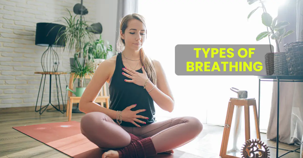
1. Diaphragmatic Breathing

Also known as belly breathing, diaphragmatic breathing involves fully engaging your diaphragm, a dome-shaped muscle located beneath your lungs. This type of breathing is often used in meditation and yoga because it helps reduce stress and promotes relaxation.

How to Practice:

  • Sit or lie down in a comfortable position.
  • Place one hand on your chest and the other on your belly.
  • Inhale deeply through your nose, ensuring your belly rises while your chest remains still.
  • Exhale slowly through your mouth.

Benefits:

  • Reduces stress and anxiety
  • Improves oxygen exchange
  • Strengthens the diaphragm

2. Thoracic Breathing

Thoracic or chest breathing involves using the muscles in your chest rather than your diaphragm. This is a more shallow form of breathing and is often the default during stressful situations.

How to Practice:

  • Sit upright and place your hand on your chest.
  • Take short, quick breaths in and out through your mouth.

Benefits:

  • Useful for quick bursts of energy
  • Helps during physical activity

However, relying solely on thoracic breathing can lead to shallow breathing habits, which may increase stress over time.

3. Nasal Breathing

Breathing through your nose is considered more efficient and healthier than mouth breathing. Your nasal passages filter, warm, and humidify the air you inhale, protecting your lungs and improving oxygen absorption.

How to Practice:

  • Keep your mouth closed.
  • Inhale and exhale gently through your nose.

Benefits:

  • Enhances oxygen uptake
  • Supports cardiovascular health
  • Reduces snoring and improves sleep quality

4. Mouth Breathing

Although not ideal for regular practice, mouth breathing can be useful in certain situations, such as during intense physical activity when your body requires more oxygen.

Drawbacks:

  • Can dry out the mouth and throat
  • May increase the risk of sleep disorders, like sleep apnea

Tip: Use mouth breathing sparingly and focus on nasal breathing whenever possible.

5. Pursed-Lip Breathing

This technique is commonly recommended for people with lung conditions like chronic obstructive pulmonary disease (COPD). It helps regulate breathing and makes it easier to exhale completely.

How to Practice:

  • Inhale through your nose for about two seconds.
  • Pucker your lips as if you’re about to whistle.
  • Exhale slowly and gently through your pursed lips for four seconds.

Benefits:

  • Improves airflow
  • Strengthens lung function

6. Box Breathing

Box breathing, also known as square breathing, is a structured breathing technique used by athletes and Navy SEALs to calm the mind and enhance focus.

How to Practice:

  • Inhale through your nose for four counts.
  • Hold your breath for four counts.
  • Exhale through your mouth for four counts.
  • Hold your breath again for four counts.

Benefits:

  • Reduces stress and improves focus
  • Enhances lung capacity
  • Promotes mental clarity

7. Yogic Breathing (Pranayama)

Pranayama is a series of breathing exercises practiced in yoga to balance the mind, body, and spirit. Techniques like alternate nostril breathing and Kapalabhati breathing fall under this category.

How to Practice (Alternate Nostril Breathing):

  • Sit comfortably and close your right nostril with your thumb.
  • Inhale deeply through your left nostril.
  • Close your left nostril with your ring finger and exhale through your right nostril.
  • Repeat the cycle for 5-10 minutes.

Benefits:

  • Balances energy levels
  • Boosts mental clarity
  • Relieves stress

Conclusion

Understanding and practicing different types of breathing can significantly enhance your health and well-being. Whether you’re looking to reduce stress, improve focus, or enhance physical performance, there’s a breathing technique for every need. Start incorporating these methods into your daily routine and feel the transformative power of mindful breathing.

Difference between Hot Pack and Cold pack in Physiotherapy

Difference between Hot Pack and Cold pack in Physiotherapy

HOT PACK

It is a kind of device wrapped in a form of Bag made up of different materials. When heat is applied to the affected area it reduces pain and promotes relaxation. It is used for muscle pain, back pain, strains, muscle stiffness and to increase blood flow. This therapy using hot packs is applied directly on the injured or exposed body area that needs treatment.

WHEN TO USE HOT PACK

1- Muscle Recovery and chronic joint pain

2- Chronic Pain and Relaxation

3- Muscle Stiffness

4- Improving mobility

Benefits of HOT PACK

1- Improves tissue Flexibility and blood circulation

2- It decreases pain in muscles

3- Heat in HOT PACK reduces pain sensitivity in joints and muscles.

4- It relaxes tight muscles which is helpful during muscle cramps.

COLD PACK

It is a therapy device made up of frozen ice. It is commonly used in injured areas to reduce pain and swelling. It is a temporary pain relief technique. It protects the tissues from further damage by slowing down blood flow inflammation.

WHEN TO USE COLD PACK

1-During Nerve and Migraine Pain

2-During Arthritis and chronic pain

3-During Sports related activity which usually causes muscle injuries or soreness

4-Insect Bites which causes itching and swelling

Benefits of COLD PACK

1- Reducing chronic pain

2- Reduces Nerve activity due to anesthesia nature of cold packs which help in severe pain

3- Treats sports injuries like sprained ankles or muscle strains and inflammation caused by it. Also reduces muscle soreness.

4- Reduces Muscles stiffness

When to use HOT and COLD Pack

Both the therapy techniques are used for injury treatment and pain relief, But are used in different conditions. Cold therapy is used to control pain immediately after an injury due to reduction in blood flow which helps swelling and inflammation. On the other hand

HOT therapy is used where the goal is to increase blood flow and relax muscles by providing heat to the inflated area. Heat can soothe the joints, cramps and abdominal muscle pain.

“At Arunalaya Healthcare, we pride ourselves on being the best physiotherapy center in Delhi. Our dedicated team of experts offers top-notch physiotherapy treatment tailored to your needs. Experience the difference with the leading physiotherapy clinic in Delhi area. Our commitment to excellence ensures that you receive the best physiotherapy care possible. Trust Arunalaya Healthcare for the best physiotherapy treatment in Delhi. Our advanced physiotherapy solutions set us apart as the premier choice for your rehabilitation needs. When it comes to physiotherapy, our center stands out as the best in Delhi. Choose Arunalaya Healthcare for comprehensive physiotherapy solutions that deliver results. Visit Arunalaya Healthcare today and discover why we are the best physiotherapy center in Delhi.”

Osteoarthritis

Osteoarthritis: Causes, Symptoms, & Physiotherapy Management

Introduction

Osteoarthritis is a degenerative joint disease, in which there is a wear and tear of cartilage in the joint. It is the most common type of arthritis and is more common in older people.

People with osteoarthritis usually have joint pain and, after rest or inactivity, stiffness for a short period of time. 

Causes

Osteoarthritis can occur due to multiple reasons. Causes can be primary or secondary –

Primary osteoarthritis

Primary osteoarthritis is also called idiopathic osteoarthritis. But even though the exact cause remains unknown, researchers have identified some risk factors that increase the probability of developing OA.

Risk factors include :-

Increasing age

Genetics

Obesity

Previous fracture

Repetitive stress

Occupations involving heavy manual work

High impact sports

Secondary osteoarthritis

Happens when something directly damages one of your joints enough to cause osteoarthritis. Injuries and traumas are common causes of secondary osteoarthritis. Other types of arthritis can damage the cartilage in the joints.

What are osteoarthritis symptoms?

The most common symptoms of osteoarthritis include:

Pain in a joint

Stiffness.

Swelling near a joint.

A decreased range of motion

A joint looking noticeably different than it used to (joint deformity).

Is osteoarthritis curable?

There’s no cure for osteoarthritis, but the condition does not necessarily get any worse over time. There are a number of treatments to help relieve the symptoms. The main treatments for the symptoms of osteoarthritis include: Physiotherapy and lifestyle measures – such as maintaining a healthy weight and exercising regularly.

Physiotherapy management includes

Manual therapy – Manual therapy helps to relieve tension from the muscles and enhance the mobility of the joint.

Deep tissue release and trigger point therapy helps to release the trigger points in the muscle in order to improve the blood flow making the muscles more efficient.

Ultrasonic and LASER therapy – US and LASER helps to promote healing of the joints and muscles thereby reducing inflammation and pain.

Strengthening exercises – Strengthening exercises plays a vital role in the management of OA as it strengthen the muscle and slows down the degenerative process.

“At Arunalaya Healthcare, we pride ourselves on being the best physiotherapy center in Delhi. Our dedicated team of experts offers top-notch physiotherapy treatment tailored to your needs. Experience the difference with the leading physiotherapy clinic in Delhi area. Our commitment to excellence ensures that you receive the best physiotherapy care possible. Trust Arunalaya Healthcare for the best physiotherapy treatment in Delhi. Our advanced physiotherapy solutions set us apart as the premier choice for your rehabilitation needs. When it comes to physiotherapy, our center stands out as the best in Delhi. Choose Arunalaya Healthcare for comprehensive physiotherapy solutions that deliver results. Visit Arunalaya Healthcare today and discover why we are the best physiotherapy center in Delhi.”
 

Benefits of physiotherapy in athletic recovery

Benefits of physiotherapy in athletic recovery

1. Prevention

Screening and assessment- therapist conduct pre participation examination to check whether an athlete is on risk of injury or not.

Education- therapist give proper info about the training, warm up, cool down to the athletes.

2. Acute care

Immediate care- therapist provide immediate management to the acute care to reduce swelling ad inflammation. Such as cryotherapy, compression, elevation, etc.

Early rehabilitation- in initial stage must focus on pain management, reduce swelling and mobility.

3. Rehabilitation

Progressive exercise therapy- rehab program must be design in a progressive way of improvement to restore strength, ranges, flexibility and functional abilty.

Pain reduces techniques

Sports massage and joint mobilization reduce joint stiffness and promote blood flow.

Some modalities like US and TENS also help in reducing the pain.

5. Neuromuscular re-education

It increases movement pattern and coordination in athletes. It helps in improving balance, proprioception and agility of an athlete.

6. Hydration

Therapist must advise for proper hydration to the athlete as dehydration cause heat stroke, heat illness and cramping to the athlete.

7. Specialised techniques

  • MFR- therapist must release the tightened muscles to gain the full range and flexibility during the play.
  • Taping- therapist must tape the weak or injured muscles. k tape used for support and inhibition and activation while rigid tape used for proving support to the instable joint and provide stability.
  • Needling – it is done to get rid of trigger points develop due to overuse of muscles.
  • Manual Therapy: Techniques like stretching, soft tissue massage, and joint mobilization help to make joints work better, ease pain, and support the healing of tissues. o Electrotherapy: Methods such as ultrasound and TENS (transcutaneous electrical nerve stimulation) along with EMS (electrical muscle stimulation) are used to heal, alleviate pain, and boost muscle function.
  • ART: Active Release Techniques (ART) are a soft tissue method that focuses on relieving tissue tension via the removal of fibrosis/adhesions which can develop in tissues as a result of overload due to repetitive use.
  • MET: This technique focuses on pain relief, increase range of motion, and strengthen the weak muscles.
Foot Drop

Foot Drop: Causes, Symptoms, and Physiotherapy Treatments

Introduction

Foot drop is a neurological condition that significantly impacts an individual’s ability to lift the front part of the foot. This condition often arises from damage or dysfunction in the nerves and muscles responsible for dorsiflexion (lifting the foot upwards). Most commonly, it is associated with injury to the common peroneal nerve, leading to weakness in the tibialis anterior muscle and other dorsiflexors. Foot drop can affect walking patterns and overall mobility, but timely intervention can help improve function and quality of life.

Causes of Foot Drop

Foot drop may stem from a variety of underlying factors, including:

  1. Nerve Damage or Compression: Injury to the common peroneal nerve, often due to prolonged pressure or trauma, can disrupt the signals required for foot movement.
  2. Muscle Weakness or Paralysis: Conditions affecting the muscles responsible for dorsiflexion, such as muscular dystrophy, may result in foot drop.
  3. Neurological Disorders: Diseases like multiple sclerosis, stroke, or amyotrophic lateral sclerosis (ALS) can impair the nervous system’s ability to control foot movement.
  4. Trauma or Injury: Direct injuries to the leg or foot can damage muscles, tendons, or nerves, leading to foot drop.
  5. Certain Medical Conditions: Diabetes and other metabolic disorders may contribute to nerve damage, increasing the risk of foot drop.

Symptoms of Foot Drop

Foot drop manifests through noticeable physical symptoms, including:

  • Difficulty Lifting the Foot: Individuals may struggle to raise the front part of their foot, causing a dragging sensation while walking.
  • Dragging the Foot: The foot may drag on the ground, leading to uneven walking patterns.
  • Slapping the Foot: Affected individuals often slap the foot down forcefully with each step.
  • High-Stepping Gait: To compensate, they may lift the thigh higher than usual while walking.
  • Pain, Numbness, or Weakness: Some may experience sensory changes or muscle weakness in the foot.

Physiotherapy Treatment for Foot Drop

Physiotherapy plays a crucial role in managing foot drop by addressing muscle weakness, nerve dysfunction, and mobility issues. Treatment may include:

1. Muscle Strengthening Exercises

  • Ankle Dorsiflexion Strengthening: Exercises targeting the tibialis anterior and other foot-lifting muscles.
  • Ankle Mobilization: Active and passive movements to enhance ankle mobility.
  • Toe Mobilization: Gentle exercises to improve flexibility and range of motion.

2. Stretching Techniques

  • Calf Muscle Stretching: Lengthening the gastrocnemius and soleus muscles to improve ankle mobility and reduce tension.

3. Trigger Point Release

  • Gastrocnemius and Soleus Release: Applying sustained pressure to specific trigger points to alleviate tension in the calf muscles.
  • Tibialis Anterior Release: Releasing tightness in the muscle responsible for ankle dorsiflexion.

4. Neuromuscular Electrical Stimulation (NMES)

Electrical impulses stimulate muscle contraction, improving strength and helping restore function.

5. Dry Needling

Dry needling helps relax tight muscles in the lower leg and foot, enhancing mobility and muscle activation for improved dorsiflexion.

6. Taping Techniques

  • Supportive Taping: Kinesiology tape applied around the ankle provides stability and promotes proper alignment.
  • Dorsiflexion Assist Taping: Tape applied from the toes to the ankle assists in lifting the foot, making walking easier.

Recommended Exercises

Specific exercises can strengthen the affected muscles and improve coordination:

  • Ankle Dorsiflexion/Plantar Flexion
  • Ankle Circles
  • Toe Raises
  • Heel Walks
  • Resistance Band Exercises

Rehabilitation Techniques

Rehabilitation focuses on restoring functional mobility and includes:

  • Walking Training: Practicing proper gait mechanics.
  • Step-Ups: Strengthening the lower limb muscles.
  • Balance Exercises: Enhancing stability with activities like single-leg stands.
  • Stretching: Achilles tendon and gastrocnemius stretches to improve flexibility.

Conclusion

Foot drop is a challenging condition, but with timely and consistent physiotherapy, individuals can regain strength, mobility, and confidence. From targeted exercises to advanced techniques like NMES and dry needling, physiotherapy offers a comprehensive approach to improving function and minimizing the impact of foot drop on daily life. If you’re experiencing symptoms of foot drop, consult a qualified physiotherapist to develop a personalized treatment plan tailored to your needs.

flat feet

Flat Feet: Causes, Classification, and Impact

What is Flat Foot?

Flat foot, medically known as pes planus, refers to a condition where the entire sole of the foot makes complete contact with the ground while standing. This occurs due to a loss of the medial longitudinal arch, a crucial structure in the foot that provides support, absorbs weight-bearing forces, and stores energy during movement.

Causes of Flat Feet

Flat feet often develop in childhood, as the arches may not fully form. However, other contributing factors include –

  • Injuries: Trauma to the foot can lead to a collapse of the arch.
  • Aging: Wear and tear over time can weaken the foot’s supportive structures.
  • Underlying Conditions: Obesity, tight calf muscles, or ligament laxity may increase the risk of developing flat feet.

Anatomy of the Medial Longitudinal Arch

The medial longitudinal arch consists of several bones, including the calcaneus, navicular, talus, cuneiforms, and the first three metatarsals. It is supported by –

  • Soft Tissues: Plantar calcaneonavicular ligament (spring ligament), deltoid ligament, and plantar aponeurosis.
  • Muscles: Posterior tibial tendon and flexor hallucis longus and brevis.

These structures work together to stabilize the arch and enable efficient foot function during walking or running.

Pathophysiology of Flat Feet

When the supporting structures of the arch are compromised, the medial longitudinal arch collapses. Factors contributing to this include:

  • Excessive tension in the triceps surae (calf muscles)
  • Posterior tibial tendon dysfunction
  • Ligament laxity in the spring ligament or plantar fascia
  • Tight Achilles tendon or calf muscle

Classification of Flat Feet

Flat feet can be classified based on two primary factors:

1. Arch Height

  • The ratio of navicular height to foot length helps determine the structure of the arch. Children’s foot arches typically develop with age, with flatness reducing over time.

2. Heel Eversion Angle

  • The heel’s eversion angle indicates foot posture. A resting calcaneal stance position can guide clinicians in assessing this posture. By age seven, the optimal vertical heel position is often achieved.

Types of Flat Feet

Flat feet are also categorized as rigid or flexible:

  • Flexible Flat Foot: The arch is visible when the foot is not bearing weight but disappears under full weight-bearing conditions.
  • Rigid Flat Foot: The arch is absent regardless of weight-bearing.

Symptoms of Flat Feet

Flat feet may be asymptomatic, but in some cases, they can alter the biomechanics of the lower body, leading to discomfort or injury.

In Children:

  • Generally asymptomatic, but some may experience pain in the arch or night cramps during adolescence.

In Adults:

  • Frequent ankle sprains or “rolling” of the ankle.
  • Pain in the midfoot, heel, lower leg, knee, hip, or back due to strained muscles and tissues.
  • Altered gait patterns in advanced cases.

Conclusion

Flat feet, while common, can range from being asymptomatic to causing significant discomfort and biomechanical issues. Understanding the causes, classification, and symptoms of pes planus can help in early diagnosis and effective management, ensuring better mobility and quality of life.

“At Arunalaya Healthcare, we pride ourselves on being the best physiotherapy center in Delhi. Our dedicated team of experts offers top-notch physiotherapy treatment tailored to your needs. Experience the difference with the leading physiotherapy clinic in Delhi area. Our commitment to excellence ensures that you receive the best physiotherapy care possible. Trust Arunalaya Healthcare for the best physiotherapy treatment in Delhi. Our advanced physiotherapy solutions set us apart as the premier choice for your rehabilitation needs. When it comes to physiotherapy, our center stands out as the best in Delhi. Choose Arunalaya Healthcare for comprehensive physiotherapy solutions that deliver results. Visit Arunalaya Healthcare today and discover why we are the best physiotherapy center in Delhi.”

brachial plexus

Brachial Plexus

The brachial plexus is a complex network of nerves that originates from the spinal cord and is crucial for the sensory and motor innervation of the upper limb. Formed by the anterior rami of spinal nerves C5 through T1, it traverses from the neck, through the axilla, and into the arm, providing essential nerve supply to the shoulder, arm, forearm, and hand.

Anatomy of the Brachial Plexus

Structure

The brachial plexus is anatomically divided into five main components: roots, trunks, divisions, cords, and terminal branches. This hierarchical organization allows for efficient distribution of nerve fibres:

  • Roots: The roots consist of five anterior rami from spinal nerves C5, C6, C7, C8, and T1. These roots converge to form three trunks.
  • Trunks: The three trunks are:
    • Superior trunk (C5-C6)
    • Middle trunk (C7)
    • Inferior trunk (C8-T1)

Divisions: Each trunk splits into an anterior and a posterior division, resulting in six divisions in total.

Cords: The divisions regroup to form three cords:

Lateral cord

Medial cord

Posterior cord

Terminal Branches: From these cords emerge five primary terminal branches that innervate various muscles and skin regions:

  • Musculocutaneous nerve
  • Median nerve
  • Ulnar nerve
  • Axillary nerve

Radial nerve

Function

The brachial plexus is responsible for both motor control and sensory perception in the upper extremity. It innervates muscles such as the biceps brachii and triceps brachii while also providing sensory input from the skin of the arm and hand. The intricate branching allows for precise control over complex movements and sensations.

Clinical Significance

Injuries to the brachial plexus can lead to significant functional impairments. These injuries are often categorized based on their location:

  • Upper Trunk Injuries (Erb’s Palsy): Typically result from trauma that stretches or tears the upper roots (C5-C6). Symptoms may include weakness in shoulder abduction and external rotation.
  • Lower Trunk Injuries (Klumpke’s Palsy): Affect the lower roots (C8-T1) and can result in weakness or paralysis of hand muscles, often associated with claw hand deformity.

Causes of Injury

Brachial plexus injuries can occur due to various traumatic events such as:  

  • Motor vehicle accidents
  • Falls
  • Sports injuries
  • Difficult childbirth

These injuries can range from mild neuropraxia, which may heal spontaneously, to severe avulsions requiring surgical intervention.

What are the common injuries to the brachial plexus

Common injuries to the brachial plexus can arise from various traumatic events, leading to significant functional impairments in the upper limb. These injuries are categorized based on their mechanism and severity, which can vary widely.

Types of Brachial Plexus Injuries

  1. Neuropraxia: This is the mildest form of injury where the nerve is stretched but not torn. It typically results in temporary symptoms such as weakness or numbness, often resolving on its own.
  2. Neuroma: In this case, the nerve has torn and healed, but scar tissue forms around it, causing pressure that impairs nerve function.
  3. Rupture: A rupture occurs when the nerve is torn but remains attached at the spinal cord. This type of injury can lead to significant loss of function and may require surgical intervention.
  4. Avulsion: The most severe form of brachial plexus injury, avulsion occurs when the nerve is completely torn from the spinal cord. This often results in debilitating pain and loss of movement in the affected arm and may necessitate complex surgical repair.

Mechanisms of Injury

Brachial plexus injuries can be caused by various mechanisms:

  • Traction Injuries: These occur when the arm is forcefully pulled away from the body, often seen in falls or during contact sports. The head may be pushed away from the shoulder, stretching the nerves.
  • Heavy Impact: Injuries can also result from direct trauma, such as a heavy blow to the shoulder or a motor vehicle accident. This impact can cause fractures or dislocations that compress or damage the brachial plexus.
  • Obstetric Injuries: During childbirth, excessive pulling on a baby’s head can lead to brachial plexus injuries, commonly referred to as Erb’s Palsy (upper trunk injury) or Klumpke’s Palsy (lower trunk injury). These injuries may affect arm movement and function.

Specific Conditions

  • Erb’s Palsy: This condition results from injury to the upper trunk (C5-C6) of the brachial plexus, often leading to weakness in shoulder abduction and external rotation. It is frequently associated with birth injuries or falls onto an outstretched arm.
  • Klumpke’s Palsy: Involves damage to the lower trunk (C8-T1) and typically results in weakness of hand muscles and a claw-like hand deformity. This can occur during traumatic events where excessive abduction of the arm happens.
Best exercise for lower back pain relief​

Best exercise for lower back pain relief​

Introduction

Back pain is a common condition that affects millions of people worldwide . It can range from mild ,acute pain to severe chronic pain that interferes with daily activities and reduce quality of life.

The back pain is complex structure made up of bones , muscle, ligament, and tendon that work together to provide support stability and movement

Back pain  can be caused by a variety of factors, including

  • Muscle strain or overuse
  • Poor posture
  • Herniated/ bulging disc
  • Degeneration condition like , osteoarthritis, spinal stenosis
  • Injury or trauma

Most common cause for low back pain

  1. Work posture – leaning forward in a chair while looking at a computer puts 3 to 4 times more strain on the back as compared to sitting upright.
  2. Abnormal load on the spine – carrying loads multiple times of your own body weight over a long period of time leads to damage to joint muscle and ultimately the pulpous discs that leads to a disc prolapse .
  3. Obesity – additional fat around the abdomen alter the spinal curves , causing exaggeration of the lower back curve ( hyperlordoisis).
  4. Genetics – very uncommonly a person may suffer from low back pain if he /she is born with a spinal defect such as scoliosis or a hemivertebrae  or a fused spine .

Physiotherapy treatment

there are various recommended physiotherapy methods of treatment –

Heat therapy-

Heat help the increase the blood circulation to the skin muscle , bones and joints. Increase blood supply takes away the pain producing sustained from the tissue and rids patient of the pain .

Cold therapy

it’s consists of ice pack, ice massage , cold water pack etc. It is very effective If used with in 24 hr. Of an acute  injury of the back. After 24-48 hr one you can switch over to heat therapy .

Manual therapy

A therapist use Their hands or elbow to apply pressure of the trigger point. trigger point release help in pain relief by releasing tension in the muscle. Releasing trigger point can help improve flexibility and range of motion . Releasing trigger point can help improve posture by reducing muscle imbalance.

IASTM

Instrument assisted soft tissue mobilization a tool is used to apply pressure to the trigger point.

Exercise for the back pain

exercises serve the role of putting spine back to its normal shape . They aimed to strengthen posterior spinal muscle , abdominal muscle and thigh ( quadriceps) muscle . To tone the other muscle of the trunk .

Abdominal muscle exercise –

1. Head raising exercise –

Best exercise for lower back pain relief​

2. Straight leg raising

exercise for lower back pain relief​

4. Knee to chest

Best exercise for lower back pain relief​

5. Abdominal flexion to toe touch

Best exercise for lower back pain relief​

6. Abdominal rotation

exercise for lower back pain relief​

Exercise of trunk hip and thigh muscle-

1. Bridging

Best exercise for lower back pain relief​

2. Trunk flexion & extension

Best exercise for lower back pain relief​

3. Hamstring stretch

Best exercise for lower back pain relief​

4. Quadriceps exercise –

Best exercise for lower back pain relief​

5. Back extensor muscle exercise

Best exercise for lower back pain relief​

Electrotherapy

Electrotherapy

Electrotherapy is a therapeutic technique that employs electrical energy to treat various medical conditions. This approach has evolved significantly since its inception, with applications spanning pain management, tissue repair, and muscle rehabilitation. This article explores the principles of electrotherapy, its various modalities, benefits, and the challenges associated with its use.

What is Electrotherapy?

Electrotherapy involves the application of electrical currents to the body to induce physiological changes. It is primarily utilized in physical therapy for:

  • Pain relief: Reducing chronic and acute pain.
  • Muscle stimulation: Enhancing muscle strength and preventing atrophy.
  • Tissue healing: Accelerating wound healing and improving circulation.
  • Neuromuscular re-education: Assisting in the recovery of motor functions post-injury or surgery.

The basic mechanism involves placing electrodes on the skin, which deliver controlled electrical impulses to targeted areas. These impulses can stimulate nerves and muscles, promoting healing and pain relief through various physiological processes.

Types of Electrotherapy Modalities

Several modalities fall under the umbrella of electrotherapy, each with unique applications:

  1. Transcutaneous Electrical Nerve Stimulation (TENS): A widely used method for pain relief that sends low-voltage electrical currents through the skin to stimulate nerves.
  2. Electrical Muscle Stimulation (EMS): Targets muscle tissues to induce contractions, beneficial for rehabilitation and preventing muscle wasting.
  3. Interferential Current Therapy (IFC): Uses two medium-frequency currents that intersect at the treatment site to reduce pain and promote healing.
  4. Ultrasound Therapy: Employs sound waves to penetrate tissues, promoting deep heating and enhancing blood flow.
  5. Microcurrent Therapy: Involves extremely low levels of electrical current to stimulate cellular repair processes.

Each of these modalities can be tailored to individual patient needs based on their specific conditions and treatment goals.

Benefits of Electrotherapy

Electrotherapy offers numerous advantages in clinical practice:

  • Non-invasive Treatment: Unlike surgical options, electrotherapy is non-invasive and generally well-tolerated by patients.
  • Pain Management: It provides an alternative to pharmacological treatments, particularly in light of the opioid crisis, offering a safer option for chronic pain management.
  • Enhanced Recovery: By improving circulation and stimulating tissue repair mechanisms, electrotherapy can accelerate recovery from injuries and surgeries.
  • Versatile Applications: It can be utilized across various medical fields, including orthopedics, neurology, and rehabilitation therapy.

Challenges and Considerations

Despite its benefits, electrotherapy is not without challenges:

Variable Effectiveness: The effectiveness of electrotherapy can vary significantly among individuals. Some patients may experience substantial relief while others may not respond at all.

Limited Evidence Base: While many practitioners advocate for its use, the scientific literature on certain modalities remains inconclusive regarding their efficacy for specific conditions.

Device Safety and Regulation: Although devices are generally deemed safe by regulatory bodies like the FDA, there are concerns about the rigorousness of approval processes compared to pharmaceuticals. Users should be cautious when purchasing over-the-counter devices without clinical guidance.

What are the latest advancements in electrotherapy

Recent advancements in electrotherapy have significantly enhanced its applications in pain management, wound healing, and rehabilitation. Here are some of the latest developments:

1. Emerging Modalities

  • Microcurrent Therapy: This technique utilizes low-level electrical currents that mimic the body’s natural bioelectric signals, promoting cellular repair and regeneration. It is increasingly recognized for its effectiveness in treating chronic pain and facilitating wound healing without device-related adverse effects.
  • Extracorporeal Shockwave Therapy (ESWT): This non-invasive treatment uses acoustic waves to promote healing in musculoskeletal conditions. Its application has expanded beyond traditional uses, showing promise in treating chronic pain syndromes and enhancing tissue repair.
  • Low-Intensity Pulsed Ultrasound (LIPUS): LIPUS is gaining traction for its ability to stimulate bone healing and soft tissue repair. It operates at lower intensities than conventional ultrasound, making it safer and more effective for sensitive conditions.

2. Innovations in Pain Management

  • Pulsed Radiofrequency Electromagnetic Therapies (PRF): This method involves high-frequency electromagnetic fields that target nerve mitochondria, leading to changes in gene expression associated with pain relief. PRF is particularly beneficial for chronic and acute pain management, often requiring only a few sessions for long-term relief.
  • Neuromuscular Electrical Stimulation (NMES): NMES continues to evolve, focusing on enhancing muscle recovery post-injury by stimulating muscle contractions. New devices are being developed to provide more targeted and effective stimulation, improving rehabilitation outcomes.

3. Applications for Wound Healing

A promising treatment for wound healing, especially for chronic lesions like diabetic ulcers, is electrostimulation therapy. Recent research shows that it can speed up the inflammatory response required for healing, increase angiogenesis, and improve oxygenation. High-voltage pulsed current (HVPC) is one technique that has demonstrated notable effectiveness in encouraging the closure of wounds that are challenging to heal.

4. Integration of Technology

The integration of advanced technologies into electrotherapy practices is becoming more common:

  • Smart Devices: New electrotherapy devices are equipped with smart technology that allows for personalized treatment plans based on real-time feedback from the patient’s response to therapy.
  • Augmented Reality (AR) and Virtual Reality (VR): These technologies are being explored to enhance patient engagement during rehabilitation exercises, making the therapy process more interactive and effective.

5. Evidence-Based Practices

    There is a growing emphasis on evidence-based approaches in electrotherapy. Research continues to refine the understanding of how different modalities can be effectively utilized across various conditions, ensuring that practitioners can provide optimal care tailored to individual patient needs.

    Duchenne Muscular Dystrophy

    Myopathies and Muscular Dystrophy: An Overview

    Introduction

    Myopathies are primary disorders of muscles that manifest as pathological, biochemical, or electrical abnormalities. These conditions are typically characterized by:

    • Proximal muscle group involvement
    • Muscle wasting
    • Absence of reflexes
    • Fibrillation
    • Bilateral involvement
    • Normal central and peripheral nervous systems
    • Progressive downhill course without remission

    Some myopathies are genetically determined and may have hereditary transmission, though their exact causes remain elusive.

    Muscular dystrophies represent a heterogeneous group of inherited disorders marked by gradual muscle fiber degeneration. Key features include:

    • Onset in early life
    • Proximal muscle involvement
    • Loss of deep tendon reflexes
    • Pseudohypertrophy of muscles

    Muscular dystrophy is distinct from other muscle diseases based on four mandatory criteria:

    1. It is a primary myopathy.
    2. There is a genetic basis for the disorder.
    3. The course is progressive.
    4. Muscle fiber degeneration and death occur at some stage of the disease.

    In muscular dystrophy, muscle wasting and weakness are symmetrical, tendon reflexes are preserved until late stages, and there is no sensory loss. The severity and progression vary significantly between types, from severe forms present at birth to slow-progressing types spanning decades.

    Classification of Muscular Dystrophies

    X-linked RecessiveAutosomal RecessiveAutosomal Dominant
    DuchenneLimb-girdle typeFacioscapulohumeral
    BeckerChildhood typeEmery-Dreifuss
    McLeod’s typeCongenital muscular dystrophyOculopharyngeal

    Duchenne Muscular Dystrophy (DMD)

    Duchenne muscular dystrophy (DMD) is the most common, extensively studied, and severe form of muscular dystrophy. It predominantly affects males, with females serving as carriers.

    Pathophysiology

    DMD is an X-linked recessive genetic disorder caused by a deletion in the DMD gene located on the X-chromosome (cytogenetic band Xp21). This gene codes for dystrophin, a protein essential for muscle membrane stabilization during contractions. The absence of dystrophin leads to muscle fiber fragility, membrane leakage, and subsequent muscular dystrophy.

    Key statistics:

    • Incidence: 1 in 3,500 live male births
    • High mutation rate: One-third of new cases arise without a prior family history
    • Dystrophin levels correlate inversely with disease severity

    Clinical Features

    Children with DMD typically exhibit muscle weakness by five years of age. Common early signs include:

    • Difficulty climbing stairs
    • Frequent falls
    • Delayed motor milestones
    • Inability to run

    Other characteristic features:

    • Onset: Between 3-6 years
    • Progressive weakness: Symmetrical and proximal
    • Pseudohypertrophy: Calf muscles and occasionally quadriceps
    • Spinal deformities: Lordosis and scoliosis
    • Gowers’ sign: A compensatory maneuver to rise from the floor due to pelvic muscle weakness
    • Cardiopulmonary involvement: Mild to moderate mental retardation

    The disease progression is predictable, transitioning through:

    1. Ambulatory stage (up to 7 years): The child can walk.
    2. Wheelchair-dependent stage (up to 12 years): Mobility declines.
    3. Bedbound stage (up to 19-20 years): Complete loss of mobility.

    Death usually occurs in the third decade due to respiratory infections or cardiomyopathy.

    Stages of DMD

    StageFunctional Ability
    Stage IFully ambulatory
    Stage IIClimbs stairs with support
    Stage IIICan stand from sitting with assistance
    Stage IVWalks with support
    Stage VWheelchair independent
    Stage VIWheelchair dependent
    Stage VIIConfined to bed (independent)
    Stage VIIIConfined to bed (dependent for all ADLs)

    Complications

    1. Respiratory

    • Recurrent infections
    • Restrictive pulmonary disease
    • Chronic alveolar hypoventilation
    • Cor pulmonale and ventricular failure

    2. Cardiomyopathy

    • Progressive heart muscle dysfunction affects both patients and carriers.

    3. Scoliosis

    • Develops during the ambulatory stage and worsens after wheelchair dependence, significantly impairing respiratory function.

    4. Contractures

    • Early contractures in the lower limbs affect mobility, while upper limb contractures result from prolonged wheelchair use.

    Investigations

    1. Serum muscle enzymes:
      • Creatine phosphokinase (CPK) levels are elevated (>10 times normal).
      • Other enzymes like pyruvate kinase and lactate dehydrogenase may also be elevated.
    2. Electromyography (EMG):
      • Detects denervated potentials in skeletal muscles.
    3. Muscle biopsy:
      • Reveals muscle degeneration and regeneration.
    4. Cardiac evaluation:
      • ECG and echocardiograms assess cardiac function.
    5. Genetic counseling:
      • Essential for family planning and carrier detection.

    Rehabilitation

    Ambulatory Stage

    • Early detection and prevention of contractures through passive stretching, prone lying, and splinting.
    • Physical therapy for endurance and respiratory muscle strengthening.
    • Psychological support for the patient and family.

    Wheelchair-Dependent Stage

    • Respiratory management, including breathing exercises and assisted ventilation.
    • Training in activities of daily living (ADL) using assistive devices.
    • Prevention of scoliosis through wheelchair modifications.

    Prolonged Survival Stage

    • Use of non-invasive respiratory aids.
    • ADL facilitation using communication aids like personal computers with voice synthesizers.

    Conclusion

    Duchenne muscular dystrophy is a devastating condition with profound physical, emotional, and psychological impacts. Early diagnosis, consistent rehabilitation, and psychological support can improve the quality of life for affected individuals and their families. Although there is no cure, advancements in genetic and supportive therapies offer hope for better management and prolonged survival. Improving awareness and providing comprehensive care remain critical in addressing the challenges of this condition.