Electrotherapy

Electrotherapy

Electrotherapy is a therapeutic technique that employs electrical energy to treat various medical conditions. This approach has evolved significantly since its inception, with applications spanning pain management, tissue repair, and muscle rehabilitation. This article explores the principles of electrotherapy, its various modalities, benefits, and the challenges associated with its use.

What is Electrotherapy?

Electrotherapy involves the application of electrical currents to the body to induce physiological changes. It is primarily utilized in physical therapy for:

  • Pain relief: Reducing chronic and acute pain.
  • Muscle stimulation: Enhancing muscle strength and preventing atrophy.
  • Tissue healing: Accelerating wound healing and improving circulation.
  • Neuromuscular re-education: Assisting in the recovery of motor functions post-injury or surgery.

The basic mechanism involves placing electrodes on the skin, which deliver controlled electrical impulses to targeted areas. These impulses can stimulate nerves and muscles, promoting healing and pain relief through various physiological processes.

Types of Electrotherapy Modalities

Several modalities fall under the umbrella of electrotherapy, each with unique applications:

  1. Transcutaneous Electrical Nerve Stimulation (TENS): A widely used method for pain relief that sends low-voltage electrical currents through the skin to stimulate nerves.
  2. Electrical Muscle Stimulation (EMS): Targets muscle tissues to induce contractions, beneficial for rehabilitation and preventing muscle wasting.
  3. Interferential Current Therapy (IFC): Uses two medium-frequency currents that intersect at the treatment site to reduce pain and promote healing.
  4. Ultrasound Therapy: Employs sound waves to penetrate tissues, promoting deep heating and enhancing blood flow.
  5. Microcurrent Therapy: Involves extremely low levels of electrical current to stimulate cellular repair processes.

Each of these modalities can be tailored to individual patient needs based on their specific conditions and treatment goals.

Benefits of Electrotherapy

Electrotherapy offers numerous advantages in clinical practice:

  • Non-invasive Treatment: Unlike surgical options, electrotherapy is non-invasive and generally well-tolerated by patients.
  • Pain Management: It provides an alternative to pharmacological treatments, particularly in light of the opioid crisis, offering a safer option for chronic pain management.
  • Enhanced Recovery: By improving circulation and stimulating tissue repair mechanisms, electrotherapy can accelerate recovery from injuries and surgeries.
  • Versatile Applications: It can be utilized across various medical fields, including orthopedics, neurology, and rehabilitation therapy.

Challenges and Considerations

Despite its benefits, electrotherapy is not without challenges:

Variable Effectiveness: The effectiveness of electrotherapy can vary significantly among individuals. Some patients may experience substantial relief while others may not respond at all.

Limited Evidence Base: While many practitioners advocate for its use, the scientific literature on certain modalities remains inconclusive regarding their efficacy for specific conditions.

Device Safety and Regulation: Although devices are generally deemed safe by regulatory bodies like the FDA, there are concerns about the rigorousness of approval processes compared to pharmaceuticals. Users should be cautious when purchasing over-the-counter devices without clinical guidance.

What are the latest advancements in electrotherapy

Recent advancements in electrotherapy have significantly enhanced its applications in pain management, wound healing, and rehabilitation. Here are some of the latest developments:

1. Emerging Modalities

  • Microcurrent Therapy: This technique utilizes low-level electrical currents that mimic the body’s natural bioelectric signals, promoting cellular repair and regeneration. It is increasingly recognized for its effectiveness in treating chronic pain and facilitating wound healing without device-related adverse effects.
  • Extracorporeal Shockwave Therapy (ESWT): This non-invasive treatment uses acoustic waves to promote healing in musculoskeletal conditions. Its application has expanded beyond traditional uses, showing promise in treating chronic pain syndromes and enhancing tissue repair.
  • Low-Intensity Pulsed Ultrasound (LIPUS): LIPUS is gaining traction for its ability to stimulate bone healing and soft tissue repair. It operates at lower intensities than conventional ultrasound, making it safer and more effective for sensitive conditions.

2. Innovations in Pain Management

  • Pulsed Radiofrequency Electromagnetic Therapies (PRF): This method involves high-frequency electromagnetic fields that target nerve mitochondria, leading to changes in gene expression associated with pain relief. PRF is particularly beneficial for chronic and acute pain management, often requiring only a few sessions for long-term relief.
  • Neuromuscular Electrical Stimulation (NMES): NMES continues to evolve, focusing on enhancing muscle recovery post-injury by stimulating muscle contractions. New devices are being developed to provide more targeted and effective stimulation, improving rehabilitation outcomes.

3. Applications for Wound Healing

A promising treatment for wound healing, especially for chronic lesions like diabetic ulcers, is electrostimulation therapy. Recent research shows that it can speed up the inflammatory response required for healing, increase angiogenesis, and improve oxygenation. High-voltage pulsed current (HVPC) is one technique that has demonstrated notable effectiveness in encouraging the closure of wounds that are challenging to heal.

4. Integration of Technology

The integration of advanced technologies into electrotherapy practices is becoming more common:

  • Smart Devices: New electrotherapy devices are equipped with smart technology that allows for personalized treatment plans based on real-time feedback from the patient’s response to therapy.
  • Augmented Reality (AR) and Virtual Reality (VR): These technologies are being explored to enhance patient engagement during rehabilitation exercises, making the therapy process more interactive and effective.

5. Evidence-Based Practices

    There is a growing emphasis on evidence-based approaches in electrotherapy. Research continues to refine the understanding of how different modalities can be effectively utilized across various conditions, ensuring that practitioners can provide optimal care tailored to individual patient needs.

    Duchenne Muscular Dystrophy

    Myopathies and Muscular Dystrophy: An Overview

    Introduction

    Myopathies are primary disorders of muscles that manifest as pathological, biochemical, or electrical abnormalities. These conditions are typically characterized by:

    • Proximal muscle group involvement
    • Muscle wasting
    • Absence of reflexes
    • Fibrillation
    • Bilateral involvement
    • Normal central and peripheral nervous systems
    • Progressive downhill course without remission

    Some myopathies are genetically determined and may have hereditary transmission, though their exact causes remain elusive.

    Muscular dystrophies represent a heterogeneous group of inherited disorders marked by gradual muscle fiber degeneration. Key features include:

    • Onset in early life
    • Proximal muscle involvement
    • Loss of deep tendon reflexes
    • Pseudohypertrophy of muscles

    Muscular dystrophy is distinct from other muscle diseases based on four mandatory criteria:

    1. It is a primary myopathy.
    2. There is a genetic basis for the disorder.
    3. The course is progressive.
    4. Muscle fiber degeneration and death occur at some stage of the disease.

    In muscular dystrophy, muscle wasting and weakness are symmetrical, tendon reflexes are preserved until late stages, and there is no sensory loss. The severity and progression vary significantly between types, from severe forms present at birth to slow-progressing types spanning decades.

    Classification of Muscular Dystrophies

    X-linked RecessiveAutosomal RecessiveAutosomal Dominant
    DuchenneLimb-girdle typeFacioscapulohumeral
    BeckerChildhood typeEmery-Dreifuss
    McLeod’s typeCongenital muscular dystrophyOculopharyngeal

    Duchenne Muscular Dystrophy (DMD)

    Duchenne muscular dystrophy (DMD) is the most common, extensively studied, and severe form of muscular dystrophy. It predominantly affects males, with females serving as carriers.

    Pathophysiology

    DMD is an X-linked recessive genetic disorder caused by a deletion in the DMD gene located on the X-chromosome (cytogenetic band Xp21). This gene codes for dystrophin, a protein essential for muscle membrane stabilization during contractions. The absence of dystrophin leads to muscle fiber fragility, membrane leakage, and subsequent muscular dystrophy.

    Key statistics:

    • Incidence: 1 in 3,500 live male births
    • High mutation rate: One-third of new cases arise without a prior family history
    • Dystrophin levels correlate inversely with disease severity

    Clinical Features

    Children with DMD typically exhibit muscle weakness by five years of age. Common early signs include:

    • Difficulty climbing stairs
    • Frequent falls
    • Delayed motor milestones
    • Inability to run

    Other characteristic features:

    • Onset: Between 3-6 years
    • Progressive weakness: Symmetrical and proximal
    • Pseudohypertrophy: Calf muscles and occasionally quadriceps
    • Spinal deformities: Lordosis and scoliosis
    • Gowers’ sign: A compensatory maneuver to rise from the floor due to pelvic muscle weakness
    • Cardiopulmonary involvement: Mild to moderate mental retardation

    The disease progression is predictable, transitioning through:

    1. Ambulatory stage (up to 7 years): The child can walk.
    2. Wheelchair-dependent stage (up to 12 years): Mobility declines.
    3. Bedbound stage (up to 19-20 years): Complete loss of mobility.

    Death usually occurs in the third decade due to respiratory infections or cardiomyopathy.

    Stages of DMD

    StageFunctional Ability
    Stage IFully ambulatory
    Stage IIClimbs stairs with support
    Stage IIICan stand from sitting with assistance
    Stage IVWalks with support
    Stage VWheelchair independent
    Stage VIWheelchair dependent
    Stage VIIConfined to bed (independent)
    Stage VIIIConfined to bed (dependent for all ADLs)

    Complications

    1. Respiratory

    • Recurrent infections
    • Restrictive pulmonary disease
    • Chronic alveolar hypoventilation
    • Cor pulmonale and ventricular failure

    2. Cardiomyopathy

    • Progressive heart muscle dysfunction affects both patients and carriers.

    3. Scoliosis

    • Develops during the ambulatory stage and worsens after wheelchair dependence, significantly impairing respiratory function.

    4. Contractures

    • Early contractures in the lower limbs affect mobility, while upper limb contractures result from prolonged wheelchair use.

    Investigations

    1. Serum muscle enzymes:
      • Creatine phosphokinase (CPK) levels are elevated (>10 times normal).
      • Other enzymes like pyruvate kinase and lactate dehydrogenase may also be elevated.
    2. Electromyography (EMG):
      • Detects denervated potentials in skeletal muscles.
    3. Muscle biopsy:
      • Reveals muscle degeneration and regeneration.
    4. Cardiac evaluation:
      • ECG and echocardiograms assess cardiac function.
    5. Genetic counseling:
      • Essential for family planning and carrier detection.

    Rehabilitation

    Ambulatory Stage

    • Early detection and prevention of contractures through passive stretching, prone lying, and splinting.
    • Physical therapy for endurance and respiratory muscle strengthening.
    • Psychological support for the patient and family.

    Wheelchair-Dependent Stage

    • Respiratory management, including breathing exercises and assisted ventilation.
    • Training in activities of daily living (ADL) using assistive devices.
    • Prevention of scoliosis through wheelchair modifications.

    Prolonged Survival Stage

    • Use of non-invasive respiratory aids.
    • ADL facilitation using communication aids like personal computers with voice synthesizers.

    Conclusion

    Duchenne muscular dystrophy is a devastating condition with profound physical, emotional, and psychological impacts. Early diagnosis, consistent rehabilitation, and psychological support can improve the quality of life for affected individuals and their families. Although there is no cure, advancements in genetic and supportive therapies offer hope for better management and prolonged survival. Improving awareness and providing comprehensive care remain critical in addressing the challenges of this condition.

    OSTEITIS PUBIS

    OSTEITIS PUBIS

    INTRODUCTION

    • Osteitis pubis is a painful chronic overuse condition affecting the pubic symphysis and surrounding soft tissues. It is characterized by pelvic pain and local tenderness over the pubic symphysis.
    • The term refers to a noninfectious, inflammatory overuse syndrome affecting the symphysis pubis and surrounding soft tissues such as musculature and fascia mainly in subjects participating in strenuous athletic activities.
    • It commonly affects athletes, especially those who participate in sports that involve kicking, turning, twisting, cutting, pivoting, sprinting, rapid acceleration and deceleration or sudden directional changes.
    • Osteitis pubis has been described in athletes who play sports such as soccer, rugby, ice hockey, Australian Rules football and distance running.
    • The diagnosis is difficult because of the anatomical complexity of the groin area, the biomechanics of the pubic symphysis region and the large number of potential sources of groin pain.

    BIOMECHANICS

    • The antagonistic relationship between the adductor longus muscle, producing an anterior-inferior force on the pubic symphysis, and the rectus abdominis muscle producing a posterior-superior force on the pubic symphysis, is particularly influential on the biomechanics of the pubic symphysis.
    • Alterations in the load-bearing characteristics of either tendon can lead to alterations in the biomechanics of the joint and instability of the pubic symphysis.
    • The anatomy of the pelvic ring is such that stresses placed on it by abnormal motion at either the sacroiliac joints or the pubic symphysis can cause instability and lead to athletic osteitis pubis.
    • The innominate bones function as arches, transferring the weight of the upright trunk from the sacrum to the hips. The fibrocartilaginous disc serves to absorb and dissipate axial and shearing forces on the pubic symphysis.
    • The mobility of the pubic symphysis under normal physiological conditions mainly involves vertical shear and anterior/posterior rotation, with resistance to bending being greater in the superior direction than the inferior direction and greater in the posterior direction than the anterior direction.
    • The relatively thick inferior pubic ligament provides the majority of joint stability in day-to-day activities, while the muscle forces acting on the pubic symphysis have considerable influence on the stress patterns in the pelvic bone.

    MECHANISM OF INJURY

    • The Centre of gravity of the body is located within the pelvis that’s why it is greatly stressed in all athletic activities.
    • The biomechanical forces are applied through the pelvis of an athlete during kicking, acceleration and deceleration which increases the incidence of osteitis pubis.
    • The shear forces produced by the repetitive contraction of the adductor muscles of the groin in the performance of sporting activities has been seen as a causative factor of instability and movement of the symphysis.
    • These forces cause chronic overloading of the pubic symphysis and parasymphyseal bone leading to a bony stress reaction.
    • Imbalances between the hip adductor and abdominal muscles have also been thought to contribute in the development of osteitis pubis.
    • Increased stress can also occur due to excessive motion. If the hip and sacroiliac joints have restricted motion, the stress of increased motion will be transferred to the pubic symphisis.
    • Direct trauma to the symphysis can also cause periostitis. Symptoms develop gradually, and might be mistaken for muscle strains.
    • Exercises that aid muscle strains might cause more irritation to the symphysis; thus, early active exercises are contraindicated.

    CLINICAL PRESENTATION

    • Anterior and medial groin pain and tenderness. Gradual onset of pain, centrally localized in the pubic symphysis with radiation to the sides and distally, is typical.
    • In some cases, pain may be centred directly over the pubic symphysis.
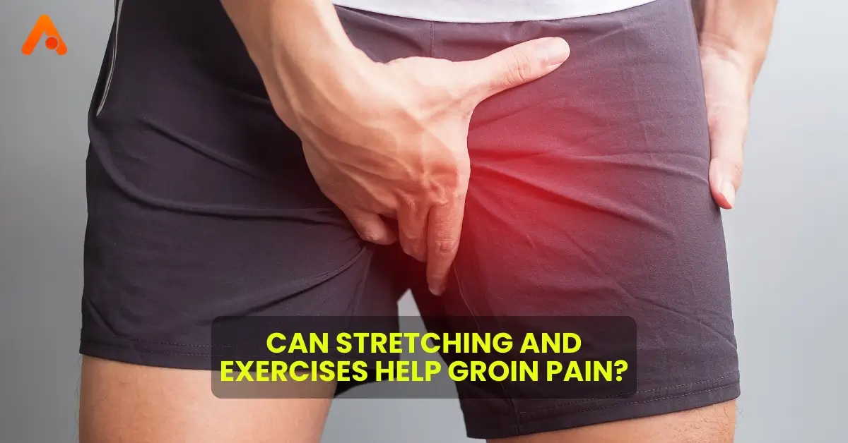
    • Pain may also be felt in the adductor region, lower abdominal muscles, perineal region, inguinal region or scrotum.
    • The pain is usually aggravated by running, cutting, hip adduction and flexion against resistance, and loading of the rectus abdominis. The pain can progress such that athletes are unable to sustain athletic activity at high levels.
    • Pain may often be more intense the morning after a training event, or when turning in bed at night.

    EXAMINATION

    • Tenderness to palpation at the symphysis, a positive adduction squeeze test, and positive hop testing.
    • ROM – There also may be restricted hip range of motion with internal and external rotation, as well as positive flexion abduction and external rotation (FABER) testing.
    • Gait Assessment – In severe cases waddling gait may be present.
    • Osteitis Pubis often is accompanied by concomitant pathology, including sacroiliac joint dysfunction, athletic pubalgia (sports hernia), femoroacetabular impingement (FAI), acetabular labral tear, chronic iliopsoas and adductor tendinopathy, and weakness in the core and pelvic stabilizer muscles making it difficult to diagnose.

    DIFFERENTIAL DIAGNOSIS

    1. Inguinal Hernia (intestines protrudes from the abdominal cavity)
      1. Pain or discomfort in the groin, especially when lifting, coughing, or bending.
    2. Sports Hernia or Athletic Pubalgia
      1. Athletic pubalgia is a disabling lower abdominal and inguinal pain possibly resulting from a hyperextension injury to the rectus insertion on the pubic symphysis, destabilizing the anterior pelvis.
      2. Insidious onset of pain with activity, resolving with rest, and radiating into the adductor, perineum, rectus, inguinal ligament, or testicular areas.
      3. Aggravated by sudden movements—coughing, sneezing, sit-ups, sprints, or kicking—ultimately limiting their performance.
    3. Femoro-Acetabular Impingement
      1. Cam and pincer lesions of the hip on X-ray.
      2. Sharp anterior pain with deep flexion, internal rotation, or abduction.
    4. Acetabular Labral Tear
      1. Combination of dull and sharp groin pain, occasionally with associated buttock pain.
      2. Symptoms worsen with activity, walking, and prolonged sitting.
      3. May be associated catching or painful clicking (mechanical symptoms)
    5. Adductor or Iliopsoas Injury
      1. Pain in the inner thigh and tenderness along the muscle belly, tendon or insertion
    6. Stress fracture – Pelvic and femoral
      1. There may be no specific findings on physical examination other than diffuse radiating pain localized to the hip or groin.
    7. Lumbar spine referred pain
    8. Ilioinguinal neuralgia (nerve entrapment syndrome)
      1. Burning and/or shooting pain that may be exacerbated by hyperextension of the hip caused by intense abdominal muscle training, direct trauma, or various inflammatory conditions.
    9. Osteomyelitis can be considered but less common cause in athletes.
      1. Local tenderness and swelling, a high temperature, occasionally an elevated erythrocyte sedimentation rate and leucocytosis.

    PT MANAGEMENT

    • Aim – to correct imbalances of the muscles acting upon the pubic symphysis, the alleged cause of Osteitis Pubis.
    • Initial treatment consists of rest from sport and reduction in weight bearing activity, followed by a progressive rehabilitation program that focuses on improving range of motion in the hips. It is postulated that this increased flexibility reduces the abnormally high biomechanical stress on the pubic symphysis.
    • Once flexibility has been achieved satisfactorily, a strengthening program should be undertaken. This should emphasize strengthening the hip adductors, abductors, and the lower abdominal musculature.
    • Consists of progressive therapeutic exercise program/ multimodal rehabilitation program is prescribed. In this program, patients are moved through different protocol stages after they are able to exercise without pain. This involves stretching, pelvic musculature strengthening, physical agents and a progressive running programme.
    • Return to sport is on average possible after 4-5 months. Occasionally symptoms may last more than 2 years.
    • Stage 1(4-6 weeks): This stage focuses on pain control and improving lumbo-pelvic stability. RICE protocol along with alternate cold (12º-14º) and hot water (31º) baths are advised. Static isometric strength exercises of the pelvic floor, of the transversus abdominis, multifidus and diaphragm, and of the adductors, and abdominal wall muscles can be started, allowing only light concentric exercises of these muscle groups, avoiding high intensity or high volume workouts. Stretching is performed except adductor and ischiopubic muscles. Cycling on an exercise bike is also started for 10mins a day and can be increased gradually.
    • Stage 2(6-8 weeks): It involves using a swiss ball for resistance and strengthening exercises of the pelvis, abdominal, and gluteal muscles.In the absence of symptoms, patients start with a maximum 30 minutes program alternating fast walk, running, and walking. Cycling on exercise bike can be increased to 20 mins a day. Changes of speed and resistance are introduced, and dynamic strengthening of the muscles around the hip in flexion, extension, adduction, and abduction are permitted by using an elastic band.
    • Stage 3 and 4(4-5 months): These include eccentric hip exercise, lunges, squats, and side-stepping with bands are done. Running is increased that involves a change of direction and pace. Kicking is only allowed at the end of the stage. Most athletes return to the pre-injury level within 3 months(4 to 14 weeks) while successful follow-up may take 6 to 14 months.
    Ultrasound physiotherapy

    Ultrasound physiotherapy

    Ultrasound physiotherapy is a specialized therapeutic technique that employs high-frequency sound waves to promote healing, reduce pain, and enhance tissue recovery. This non-invasive modality has gained popularity in the field of physical therapy due to its effectiveness in treating various musculoskeletal conditions. This article delves into the mechanisms of ultrasound therapy, its benefits, applications, and considerations for patients.

    How Ultrasound Physiotherapy Works

    Ultrasound therapy utilizes a device known as an ultrasound machine, which consists of a transducer that emits sound waves at frequencies typically between 1 and 3 megahertz. These sound waves penetrate the skin and interact with underlying tissues, leading to two primary effects-

    • Thermal Effects: The ultrasound waves generate heat within the tissues. This thermal effect increases blood flow, reduces muscle tension, and promotes relaxation in the affected area. It is particularly beneficial for chronic pain conditions and muscle spasms.
    • Non-Thermal Effects: Ultrasound also produces mechanical effects through cavitation and acoustic streaming. Cavitation involves the formation and collapse of tiny gas bubbles in the tissue fluids, which can aid in tissue repair and reduce inflammation. Acoustic streaming enhances fluid movement within tissues, facilitating nutrient exchange and waste removal.

    Benefits of Ultrasound Physiotherapy

    Ultrasound therapy offers several advantages for patients undergoing rehabilitation:

    1. Pain Relief: One of the most significant benefits is its ability to alleviate pain associated with various conditions such as arthritis, tendonitis, and muscle strains. The heating effect can help diminish discomfort by relaxing tight muscles and reducing spasms.
    2. Enhanced Healing: By increasing blood flow to the injured area, ultrasound promotes faster healing of tissues. This is particularly useful for soft tissue injuries, surgical wounds, and chronic conditions.
    3. Reduction of Inflammation: Ultrasound therapy can help reduce swelling and inflammation around injured areas by promoting lymphatic drainage and improving circulation.
    4. Scar Tissue Management: The mechanical effects of ultrasound can break down scar tissue formations, improving flexibility and range of motion in affected joints or muscles.
    5. Improved Tissue Mobility: The thermal effects allow for better stretching of muscles and tendons, which can enhance overall mobility during rehabilitation exercises.

    Applications of Ultrasound Physiotherapy

    Ultrasound physiotherapy is versatile and can be applied to various conditions, including:

    • Musculoskeletal Injuries: Effective for treating sprains, strains, tendonitis, bursitis, frozen shoulder, and other soft tissue injuries.
    • Post-Surgical Recovery: Facilitates healing after surgeries by promoting blood flow and reducing scar tissue formation.
    • Chronic Pain Conditions: Beneficial for patients suffering from chronic pain syndromes like fibromyalgia or arthritis.

    Wound Healing: Used to enhance healing in surgical wounds or chronic ulcers by promoting cellular activity and blood circulation.

    Considerations and Safety

    While ultrasound therapy is generally safe and well-tolerated, there are some considerations:

    • Contraindications: Patients with certain conditions should avoid ultrasound therapy. These include pregnancy (over the abdomen), active infections in the treatment area, malignancies near the treatment site, or implanted medical devices like pacemakers.

    Therapist Expertise: The proficiency of the physiotherapist delivering ultrasound therapy has a significant impact on its efficacy. To guarantee safety and optimize therapeutic results, proper technique is essential.
    Treatment Length: Depending on the area being treated and the particular ailment being addressed, sessions usually run five to fifteen minutes.

    How does ultrasound therapy compare to other physiotherapy techniques

    Ultrasound therapy is a widely used modality in physiotherapy that employs high-frequency sound waves to treat various musculoskeletal conditions. When comparing ultrasound therapy to other physiotherapy techniques, several key differences and similarities emerge, influencing their respective applications and effectiveness.

    Mechanisms of Action

    • Ultrasound Therapy: Utilizes sound waves to create deep tissue heating and mechanical effects, enhancing blood flow, reducing inflammation, and promoting tissue healing. The thermal effect helps relax muscles and decrease pain, while non-thermal effects can stimulate cellular activity and collagen production.
    • Electrical Stimulation (e.g., TENS): Involves sending electrical currents through the skin to stimulate nerves and muscles. This technique is primarily used for pain relief, muscle re-education, and improving circulation. Unlike ultrasound, electrical stimulation directly affects nerve pathways and can provide immediate pain relief through nerve block mechanisms.

    Ultrasound Physiotherapy Uses

    Ultrasound therapy: is frequently used to treat soft tissue injuries like bursitis, tendinitis, and the healing process following surgery. Because it may enter tissues painlessly, it works especially well for deep tissue diseases.

    Electrical Stimulation: Used for a broader range of conditions including muscle atrophy prevention, pain management in acute injuries, and rehabilitation post-surgery. It is especially beneficial for patients who require muscle strengthening or neuromuscular re-education.

    Ultrasound Physiotherapy Benefits

    Ultrasound Therapy

    • Reduces pain and muscle spasms.
    • Increases blood flow and promotes healing.
    • Breaks down scar tissue, enhancing range of motion.
    • Non-invasive with minimal side effects when administered by qualified therapists.

    Electrical Stimulation

    • Provides immediate pain relief.
    • Reduces muscle spasms and promotes muscle contraction.
    • Improves circulation and can help prevent muscle atrophy.
    • Often used in conjunction with exercise programs for enhanced rehabilitation outcomes.

    Limitations

    Ultrasound Therapy: While generally effective, its benefits can be influenced by the skill of the therapist and the specific condition being treated. Some studies suggest that the perceived benefits may include a placebo effect, highlighting the importance of using ultrasound as part of a comprehensive treatment plan rather than a standalone solution.

    Electrical Stimulation: May not be suitable for all patients, particularly those with certain medical conditions like pacemakers or skin sensitivities. Additionally, its effectiveness can vary based on electrode placement and patient tolerance to electrical impulses.

    Sports massage therapist

    Why Every Athlete Needs a Sports Massage Therapist in Their Routine

    Athletes constantly push their bodies to the limits, whether they’re training for a marathon, competing in a championship, or breaking personal records. However, this relentless pursuit of excellence often comes at a cost—tight muscles, fatigue, and an increased risk of injuries. This is where a sports massage therapist becomes essential. Incorporating regular sessions with a sports massage therapist into an athlete’s routine can enhance performance, accelerate recovery, and reduce the risk of injuries.

    What Is a Sports Massage Therapist?

    A sports massage therapist specializes in techniques that cater specifically to the needs of athletes. Unlike traditional massages that focus primarily on relaxation, sports massage therapy targets deeper layers of muscles and connective tissues. It involves a combination of stretching, compression, and trigger-point therapy to address muscle imbalances, prevent injuries, and improve overall mobility.

    Benefits of Having a Sports Massage Therapist in Your Routine

    1. Improved Performance

    A sports massage therapist helps athletes maintain peak performance by enhancing blood flow, improving flexibility, and reducing muscle tension. These factors contribute to better endurance, agility, and strength, which are essential for athletic success.

    2. Faster Recovery

    After intense workouts or competitions, muscles often experience soreness and stiffness. Sports massage therapy accelerates the recovery process by reducing lactic acid buildup and increasing oxygen supply to muscles. This allows athletes to bounce back faster and perform consistently.

    3. Injury Prevention

    Overused or tight muscles can lead to injuries such as strains, sprains, and tears. Regular sessions with a sports massage therapist ensure that potential problem areas are identified and addressed early, minimizing the risk of serious injuries.

    4. Enhanced Mental Focus

    Athletes need more than just physical readiness—they also need mental clarity. Sports massage therapy promotes relaxation by reducing cortisol levels, which helps athletes stay focused and calm during competitions.

    5. Tailored Treatment for Specific Needs

    A professional sports massage therapist understands that every sport places different demands on the body. They customize their techniques to target specific muscle groups, ensuring that the therapy aligns perfectly with the athlete’s sport and goals.

    Techniques Used by Sports Massage Therapists

    Sports massage therapy isn’t a one-size-fits-all approach. Therapists use various techniques depending on the athlete’s needs and the phase of their training. Some common methods include:

    • Deep Tissue Massage: Targets deeper layers of muscles to alleviate chronic tension.
    • Trigger Point Therapy: Focuses on releasing tight knots in muscles.
    • Pre-Event Massage: Prepares the body for physical activity by warming up muscles and increasing blood flow.
    • Post-Event Massage: Aids recovery by reducing soreness and stiffness.
    • Stretching Techniques: Improves range of motion and flexibility.

    Why Every Athlete Should Prioritize Sports Massage Therapy

    The human body is an athlete’s most valuable asset. Investing in regular sessions with a sports massage therapist isn’t a luxury—it’s a necessity. Here’s why:

    1. Consistency Is Key: Athletes who incorporate sports massage therapy into their routine experience fewer interruptions in training due to injuries or fatigue.
    2. Long-Term Benefits: Beyond immediate recovery, consistent therapy promotes long-term muscular health and reduces the risk of overuse injuries.
    3. Competitive Edge: Athletes who prioritize their physical and mental well-being often outperform their peers.

    Finding the Right Sports Massage Therapist

    When choosing a sports massage therapist, ensure they are certified, experienced, and familiar with the demands of your sport. Clinics like Arunalaya HealthCare offer advanced therapies tailored for athletes, combining expertise with the latest technology to deliver optimal results.

    Conclusion

    For athletes striving for excellence, a sports massage therapist is not just an option—it’s an essential part of their routine. From boosting performance to preventing injuries and enhancing recovery, the benefits are undeniable. By incorporating sports massage therapy into your regimen, you can ensure your body remains resilient, agile, and ready to conquer every challenge.

    Take the first step towards optimizing your athletic journey. Book a session with a sports massage therapist at Arunalaya today and experience the difference!

    Exercises for kids with Autism

    Effective Exercises for Kids with Autism: Boost Development and Well-being

    Children with autism spectrum disorder (ASD) often face challenges in motor skills, coordination, and social interaction. Exercise plays a vital role in their physical, mental, and emotional development. Carefully planned activities can improve balance, muscle strength, and sensory integration while also being enjoyable and confidence-boosting.

    This guide will delve into effective exercises for kids with autism, providing insights into their benefits, techniques, and tips for creating a structured yet fun fitness routine.

    Why Exercise is Important for Kids with Autism

    Engaging in physical activities offers numerous benefits:

    Encourages Routine: A consistent exercise schedule fosters a sense of structure and discipline.

    Improves Motor Skills: Autism often impacts fine and gross motor abilities. Exercise helps improve coordination, balance, and strength.

    Enhances Social Skills: Group activities encourage interaction, teamwork, and communication.

    Promotes Emotional Well-being: Physical activities release endorphins, reducing anxiety and promoting a sense of calm.

    Supports Sensory Integration: Certain exercises can help regulate sensory responses, making children more comfortable in different environments.

    Effective Exercises for Kids with Autism

    1. Yoga and Stretching

    Benefits: Improves flexibility, concentration, and sensory awareness.

    Cat-Cow Stretch: Promotes sensory integration.
    Incorporate visual aids or music to make yoga engaging and interactive.

    Child’s Pose: Encourages relaxation.

    Tree Pose: Enhances balance and focus.

    2. Walking or Running

    Benefits: Boosts cardiovascular health and energy levels.

    • Start with short walks in a calm environment.
    • Gradually introduce jogging or fun runs.
    • Use incentives like walking towards a favorite location or following a trail of markers.

    3. Obstacle Courses

    Benefits: Develops motor planning and problem-solving skills.

    • Create simple courses using household items (pillows, cones, or ropes).
    • Include activities like crawling under tables, hopping over objects, and balancing on a beam.
    • Modify difficulty levels based on the child’s abilities.

    4. Swimming

    Benefits: Enhances sensory regulation, strength, and coordination.

    • Water activities can be soothing for many children with autism.
    • Focus on basic skills like floating or splashing to build comfort.
    • Always ensure safety with supervision and flotation devices.

    5. Ball Games

    Benefits: Improves hand-eye coordination and social interaction.

    • Start with simple throwing and catching.
    • Gradually introduce games like basketball or soccer.
    • Use lightweight, soft balls to ensure safety.

    6. Trampoline Jumping

    Benefits: Aids in sensory processing and energy regulation.

    • Jumping helps release pent-up energy while enhancing balance.
    • Supervise and encourage creative movement on the trampoline.

    7. Dance and Music

    Benefits: Combines movement with rhythm, improving coordination and mood.

    • Use the child’s favorite songs to create a fun dance routine.
    • Encourage free movement to foster creativity.

    8. Martial Arts

    Benefits: Boosts self-discipline, focus, and self-confidence.

    • Opt for beginner-friendly styles like karate or taekwondo.
    • Choose classes with instructors experienced in working with kids with autism.

    Tips for Implementing Exercise Routines

    • Personalize Activities: Adapt exercises to the child’s preferences and abilities.
    • Consistency is Key: Establish a regular schedule to build familiarity.
    • Incorporate Rewards: Positive reinforcement encourages participation.
    • Focus on Fun: Make sessions enjoyable rather than overly structured.
    • Monitor Progress: Celebrate achievements, no matter how small.

    Conclusion

    Regular physical activities are transformative for children with autism. They foster growth in motor skills, social interaction, and emotional well-being while helping children explore their potential in a fun and supportive way. By integrating the right exercises for kids with autism into their routines, parents and caregivers can nurture development and confidence in every child.

    At Arunalaya, we believe in holistic care, offering tailored support for children with special needs. Our team is dedicated to enhancing their quality of life through innovative therapies and guidance.

    For expert advice and support, reach out to us today and embark on a journey toward brighter possibilities.

    Laser physiotherapy

    The Ultimate Guide to Laser Physiotherapy: Transforming Pain Management and Recovery

    Introduction

    In the ever-evolving field of physiotherapy, laser therapy stands out as a groundbreaking technology. Known for its ability to relieve pain, accelerate healing, and improve mobility, laser physiotherapy is rapidly becoming a preferred choice for many patients. Whether you’re dealing with chronic pain, recovering from a sports injury, or seeking rehabilitation post-surgery, laser therapy can offer effective and non-invasive treatment options. This blog dives deep into everything you need to know about laser physiotherapy, its applications, benefits, and how it works.

    What Is Laser Physiotherapy?

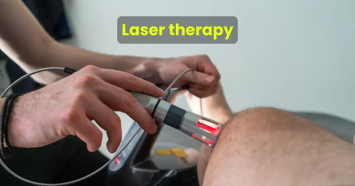
    Laser-physiotherapy uses advanced laser technology to treat various musculoskeletal conditions. The term “laser” stands for Light Amplification by Stimulated Emission of Radiation, and in physiotherapy, lasers emit specific wavelengths of light that penetrate deep into tissues. This stimulates cellular repair, reduces inflammation, and enhances blood circulation, providing therapeutic benefits.

    The procedure is performed using a laser physiotherapy machine, which is designed for safety and precision. By targeting specific areas, the therapy helps in pain relief and tissue regeneration, making it a versatile treatment option for both acute and chronic conditions.

    How Does Laser Therapy Work?

    Laser physiotherapy works on the principle of photobiomodulation. When laser light penetrates the skin, it interacts with cells, promoting the production of ATP (adenosine triphosphate), the energy currency of the cell. This process accelerates tissue repair and reduces inflammation. Here’s a breakdown of how it works:

    1. Light Absorption: The light from the laser is absorbed by chromophores in the cells.
    2. Energy Conversion: The absorbed light energy is converted into biochemical energy.
    3. Cellular Activation: This triggers cellular processes that reduce pain and inflammation and speed up healing.

    Benefits of Laser Physiotherapy

    Laser physiotherapy offers a host of benefits for patients, including:

    1. Non-Invasive Pain Relief: Ideal for conditions like arthritis, back pain, and sports injuries.
    2. Speeds Up Healing: Encourages faster tissue repair and regeneration.
    3. Reduces Inflammation: Effective in minimizing swelling and redness in affected areas.
    4. Improves Mobility: Helps restore joint and muscle function.
    5. Customizable Treatment: The intensity and duration can be adjusted for individual needs.

    Common Conditions Treated with Laser Physiotherapy

    Laser therapy is highly effective for a range of conditions, including:

    • Sports Injuries: Sprains, strains, and ligament tears.
    • Chronic Pain: Back pain, neck pain, and migraines.
    • Post-Surgical Rehabilitation: Promotes healing and reduces scar tissue.
    • Arthritis: Eases joint pain and stiffness.
    • Neurological Issues: Assists in managing nerve pain and neuropathy.

    The Role of Laser Physiotherapy Machines

    A laser physiotherapy machine is an essential tool in delivering effective treatment. These machines vary in type, including low-level lasers for superficial treatments and high-power lasers for deeper tissue therapy. Equipped with advanced features, they ensure targeted therapy with minimal side effects.

    Who Can Benefit from Laser Physiotherapy?

    Laser-physiotherapy is suitable for people of all ages and is particularly beneficial for:

    • Athletes recovering from injuries.
    • Patients with chronic conditions like osteoarthritis.
    • Individuals undergoing post-surgical rehabilitation.
    • Those seeking non-invasive alternatives to pain management.

    Is Laser Therapy Safe?

    One of the major advantages of laser therapy is its safety profile. When administered by trained professionals, such as the experts at Arunalaya, the treatment is painless and free from significant side effects. Patients often experience a warm, soothing sensation during the procedure.

    Why Choose Arunalaya for Laser Physiotherapy?

    At Arunalaya, we are committed to providing cutting-edge treatments tailored to individual needs. Our state-of-the-art laser physiotherapy machines and experienced physiotherapists ensure optimal results for every patient. Whether you’re dealing with a sports injury, chronic pain, or a neurological condition, our customized care plans prioritize your recovery and well-being.

    Conclusion

    Laser physiotherapy represents the future of non-invasive pain management and rehabilitation. By harnessing the power of laser technology, this therapy not only alleviates pain but also accelerates recovery, improves mobility, and enhances overall quality of life. If you’re considering laser-physiotherapy, consult with experts who understand your unique needs.

    At Arunalaya, we specialize in physiotherapy laser treatment using advanced tools and techniques to deliver unparalleled care. Schedule your appointment today and take the first step towards a pain-free life.

    Cervical Spondylitis

    Cervical Spondylitis: Causes, Symptoms & Treatment

    Cervical spondylitis is a chronic inflammatory condition that affects the cervical spine, causing discomfort and mobility challenges. This comprehensive guide explores cervical spondylitis in detail, including its causes, symptoms, diagnosis, treatment options, and practical self-care strategies to help manage the condition effectively.

    Introduction

    Living with cervical spondylitis can be challenging, but understanding the condition is the first step toward effective management. This guide outlines the intricacies of cervical spondylitis, offering insights into its causes, symptoms, treatment, and prevention methods.

    Understanding Cervical Spondylitis

    What is Cervical Spondylitis?

    Cervical spondylitis, also known as cervical spondylarthritis, is a condition characterized by inflammation of the vertebrae and surrounding tissues in the neck. It can result in significant pain, stiffness, and limited mobility, impacting daily activities and overall quality of life.

    Causes

    The exact cause of cervical spondylitis varies, but several factors may contribute to its development:

    • Autoimmune Response: The immune system may mistakenly attack healthy tissues in the cervical spine.
    • Genetic Predisposition: Family history increases susceptibility.
    • Environmental Triggers: Infections or physical stress can exacerbate symptoms.
    • Aging: Wear and tear on spinal structures naturally occur over time.

    Symptoms of Cervical Spondylitis

    Symptoms of cervical spondylitis can differ among individuals but commonly include:

    • Neck Pain and Stiffness: Persistent discomfort, especially during movement.
    • Radiating Pain: Pain spreading to the shoulders, arms, or hands.
    • Numbness and Tingling: Sensations caused by nerve compression.
    • Reduced Range of Motion: Difficulty turning or tilting the head.
    • Fatigue: Chronic inflammation often results in general fatigue.

    Diagnosis

    Accurate diagnosis is crucial for effective management. The diagnostic process typically includes:

    • Medical History Review: Evaluating symptoms and prior health conditions.
    • Physical Examination: Checking neck movement and identifying tender areas.
    • Imaging Tests: X-rays or MRI scans visualize inflammation and structural changes.

    Treatment Options

    Cervical spondylitis has no definitive cure, but several treatments can effectively manage symptoms.

    Medications

    • NSAIDs (Nonsteroidal Anti-inflammatory Drugs): Reduce inflammation and alleviate pain.
    • Corticosteroids: Prescribed for severe symptoms to control inflammation.

    Physical Therapy

    Regularly practicing tailored exercises can improve neck strength and flexibility while reducing stiffness.

    Lifestyle Modifications

    • Posture Correction: Maintaining good posture decreases strain on the cervical spine.
    • Regular Exercise: Low-impact activities such as walking or swimming enhance mobility.
    • Stress Management: Techniques like yoga or meditation can reduce symptom flare-ups.

    Effective Self-Care Strategies

    Self-care plays a significant role in managing cervical spondylitis and enhancing quality of life.

    1. Regular Exercise

    Incorporate physical activity to improve neck strength and flexibility:

    • Stretching: Gentle stretches increase neck mobility.
    • Strengthening: Exercises targeting neck and shoulder muscles provide better support.
    • Low-Impact Activities: Walking or swimming maintains overall fitness without added strain.

    2. Posture Awareness

    Maintaining proper posture reduces pressure on the neck:

    • Ergonomic Adjustments: Optimize workspace setup for comfort.
    • Mindful Posture: Align your head and spine while sitting or standing.

    3. Heat and Cold Therapy

    Alternating between heat and cold can relieve symptoms:

    • Heat: Use warm compresses to relax muscles and boost blood flow.
    • Cold: Apply ice packs to reduce inflammation and numb sharp pain.

    4. Pain Relief Medications

    Over-the-counter options include:

    • NSAIDs: Alleviate pain and inflammation.
    • Acetaminophen: Effective for general pain relief.

    5. Physical Therapy

    Consulting a physiotherapist can provide customized care:

    • Postural Training: Techniques to improve posture and alleviate strain.
    • Manual Therapy: Hands-on methods to enhance mobility and reduce pain.

    6. Soft Neck Brace

    Wearing a soft neck brace offers temporary support during acute pain episodes but should be used sparingly.

    7. Massage Therapy

    Relieve tension in neck and shoulder muscles through:

    • Professional Massages: Seek therapists experienced in treating cervical issues.
    • Self-Massage: Use light pressure or tools like massage balls.

    8. Alternative Therapies

    Consider complementary treatments such as:

    • Chiropractic Care: Spinal manipulation can improve function.
    • Acupuncture: Targeted treatments may alleviate pain for some individuals.

    9. Stress Management

    Chronic stress can worsen symptoms. Practice:

    • Meditation: Helps manage stress and improves focus.
    • Yoga: Gentle poses improve flexibility and strength.

    10. Healthy Lifestyle Choices

    Incorporate these habits into your routine:

    • Balanced Diet: Focus on anti-inflammatory foods rich in omega-3s and whole grains.
    • Hydration: Drink plenty of water to maintain spinal health.

    Prognosis and Lifestyle Considerations

    Cervical spondylitis is a chronic condition requiring consistent management. Untreated symptoms can severely impact quality of life, but proactive care ensures better outcomes.

    Prevention Strategies

    To reduce the risk of cervical spondylitis or minimize symptoms:

    • Maintain a healthy weight.
    • Avoid prolonged poor posture during desk work.
    • Include regular stretching and strengthening exercises in your routine.

    Conclusion

    While cervical spondylitis is a challenging condition, proper understanding, treatment, and self-care strategies can significantly improve daily life. By adopting a proactive approach and making lifestyle adjustments, individuals can effectively manage symptoms and maintain a fulfilling quality of life.

    Cervical Spine

    Cervical Spine

    Introduction

    The cervical spine plays a crucial role in supporting the head, enabling movement, and protecting the spinal cord. It consists of unique features and structures that distinguish it from other regions of the vertebral column. This article explores the anatomy, joints, ligaments, and muscles of the cervical spine while emphasizing its function and importance.

    Major Functions of the Cervical Joint

    The cervical spine performs the following vital functions-

    • Support and Cushioning: It supports and cushions loads to the head and neck while allowing for rotation.
    • Protection: It protects the spinal cord that extends from the brain.

    The cervical spine is subjected to extrinsic factors such as repetitive movements, whole-body vibrations, and static loads.

    Distinguishing Features of Cervical Vertebrae

    The cervical vertebrae are uniquely characterized by-

    • Triangular Vertebral Foramen
    • Bifid Spinous Process: The spinous process splits into two distally.
    • Transverse Foramina: These are holes in the transverse processes that provide passage to the vertebral artery, vein, and sympathetic nerves.

    Atlas (C1)

    The atlas is the first cervical vertebra and articulates with the occiput of the head and the axis (C2).

    Key Features of the Atlas:

    • Lateral Masses: Connected by anterior and posterior arches. Each lateral mass contains:
      • Superior articular facet (for articulation with occipital condyles).
      • Inferior articular facet (for articulation with C2).
    • Anterior Arch: Contains a facet for articulation with the dens of the axis, secured by the transverse ligament.
    • Posterior Arch: Contains a groove for the vertebral artery and C1 spinal nerve.

    Axis (C2)

    The axis is easily identifiable due to its dens (odontoid process), which extends superiorly from the anterior portion of the vertebra.

    Key Features of the Axis:

    • The dens articulates with the anterior arch of the atlas, creating the medial atlanto-axial joint.
    • This joint allows for the independent rotation of the head relative to the torso.

    Joints of the Cervical Spine

    The cervical spine comprises two types of joints:

    Joints Present Throughout the Vertebral Column

    1. Disc Joint:
      • Located between vertebral bodies.
      • Made of fibrocartilage (cartilaginous joint, symphysis).
      • Functions:
        • Bears the body’s weight above it.
        • Provides motion, contributing to 25% of the spine’s height and 40% of the cervical spine’s height.
    2. Facet Joint:
      • Articulation of superior and inferior articular processes from adjacent vertebrae (synovial joint).
      • Also known as zygapophyseal or Z joints.
      • Functions:
        • Guides motion at segmental joint levels.
        • The plane of the cervical facets is approximately 45 degrees, resembling a roof slope.

    Ligaments of the Cervical Spine

    Craniovertebral Ligaments

    • Anterior Atlanto-Occipital Membrane: Connects the foramen magnum to the atlas; continues with the anterior longitudinal ligament.
    • Apical Ligament: Short ligament attaching to the anterior part of the foramen magnum.
    • Alar Ligaments:
      • Inserted onto the occipital condyles.
      • Limit axial rotation between the occiput and atlas.
      • Trauma or inflammatory diseases can damage these ligaments, increasing axial rotation.
    • Membrane of Tectoria: Extends from the posterior surface of the axis body to the basiocciput.
    • Transverse Ligament of the Atlas: Secures the dens to the anterior arch of the atlas.
    • Accessory Atlanto-Axial Ligaments
    • Posterior Atlanto-Occipital Membrane
    • Lateral Atlanto-Occipital Ligaments

    Lower Cervical Ligaments

    • Anterior Longitudinal Ligament: Lies anterior to vertebral bodies; relaxed in flexion and taut in extension.
    • Posterior Longitudinal Ligament: Lies posterior to vertebral bodies in the vertebral canal; stretches in neck flexion and relaxes in extension.
    • Ligamenta Flava: Connects laminae of adjacent vertebrae; allows flexion and prevents hyper-flexion.
    • Ligamentum Nuchae:
      • A fibroelastic membrane extending from the occiput to cervical spines.
      • Provides head and neck stability, especially during flexion and acceleration injuries.

    Muscles of the Cervical Spine

    Posterior Muscles

    • Trapezius (Traps):
      • Most superficial; extends from the occiput to the lower thoracic spine.
      • Functions: Neck extensor, ipsilateral lateral flexion, and contralateral rotation.
    • Levator Scapulae:
      • Deep to traps; extends from the first four cervical vertebrae to the scapula.
      • Functions: Scapular elevation and ipsilateral lateral flexion/rotation.
    • Splenius Capitis and Cervicis: Prime movers of neck and head (extension and rotation).
    • Semispinalis Capitis and Cervicis: Deepest posterior muscles.

    Lateral Muscles

    • Scalene (Anterior, Medial, Lateral):
      • Functions: Flexion, lateral flexion, and stabilization of the cervical spine.
      • Important anatomical relationship: The brachial plexus, subclavian artery, and vein pass between the anterior and middle scalene muscles.
    • Sternocleidomastoid (SCM):
      • Extends from the manubrium and clavicle to the mastoid process.
      • Functions: Lower cervical flexion, head extension, ipsilateral lateral flexion, and contralateral rotation.

    Anterior Muscles

    • Deep Craniocervical Flexors: Longus capitis, longus colli, rectus capitis anterior, and rectus capitis lateralis.
      • Provide dynamic support to the cervical spine.
      • Work with SCM for cervical flexion.
    • Mandibular Elevator Group: Masseter, temporalis, and internal pterygoid muscles.

    Conclusion

    The cervical spine is a highly specialized structure that supports the head, enables movement, and protects the spinal cord. Its unique features, intricate joints, robust ligaments, and powerful muscles make it an essential part of the body. Understanding its anatomy and function is vital for diagnosing and treating conditions affecting the neck and upper spine.

    Kinesiology tape

    Kinesiology tape

    Introduction

    Kinesiology tape has become a widely recognized tool in physical therapy and sports medicine, praised for its ability to support muscles and joints, alleviate pain, and enhance physical performance. Developed by Dr. Kenzo Kase in the 1970s, this innovative tape mimics the skin’s elasticity, enabling natural movement while providing therapeutic benefits. Its diverse applications, ranging from injury recovery to chronic pain management, have made it a staple in rehabilitation programs worldwide.

    Key Features of Kinesiology Tape

    1. Material Composition
      Kinesiology tape is crafted from a blend of cotton and nylon, ensuring breathability and stretchiness. It adheres to the skin using a strong, water-resistant adhesive and is often available in latex-free, hypoallergenic options to suit sensitive users.
    2. Functionality
      By creating a gentle lift in the skin, kinesiology tape helps reduce pressure on underlying tissues. This unique lifting mechanism improves circulation and decreases pain signals sent to the brain, promoting faster recovery.
    3. Application Techniques
      The tape can be applied in various patterns—such as I, Y, or X—depending on the user’s needs. Proper application is essential to achieve desired outcomes, and consulting a trained professional is often recommended for optimal results.

    Benefits of Kinesiology Tape

    • Pain Relief
      Many users report decreased pain after application, as kinesiology tape interrupts pain signals and fosters a sense of relief.
    • Reduced Inflammation
      Enhanced blood flow and lymphatic drainage help mitigate swelling and inflammation in injured areas.
    • Enhanced Performance
      Athletes use kinesiology tape to support fatigued or injured muscles, enabling better performance during competition.
    • Muscle Support
      The tape stabilizes weak or injured muscles without hindering movement, facilitating improved functionality during physical activity.
    • Posture Correction
      By offering feedback through the skin, kinesiology tape aids in correcting postural issues, increasing awareness of body positioning.
    • Support for Chronic Conditions
      Its role in conditions involving muscle overuse or chronic pain, such as tendonitis or sprains, is particularly noteworthy.

    Specific Conditions Treated by Kinesiology Tape

    1. Sports Injuries
      Kinesiology tape is commonly applied for injuries like ankle sprains, shoulder impingement, and tennis elbow. It supports the injured area while allowing continued movement, aiding recovery.
    2. Chronic Pain
      Conditions such as fibromyalgia and chronic low back pain benefit from the tape’s ability to stabilize affected areas and reduce discomfort.
    3. Muscle Strains and Sprains
      Acute muscle strains and sprains are managed effectively using kinesiology tape, which alleviates pain by reducing pressure on sensitive tissues.
    4. Postural Issues
      Poor posture, a common cause of pain, can be corrected with kinesiology tape, which encourages proper alignment of the spine and shoulders.
    5. Swelling and Inflammation
      The tape’s ability to improve lymphatic drainage and circulation makes it effective in reducing swelling from conditions like lymphedema or post-surgical recovery.
    6. Joint Pain
      Arthritis and general joint pain can benefit from kinesiology tape’s supportive properties, which stabilize joints without limiting mobility.
    7. Post-Surgical Rehabilitation
      During rehabilitation after surgery, kinesiology tape helps restore mobility and function while protecting healing tissues.
    8. Trigger Points
      By lifting the skin over tense muscles, kinesiology tape relieves pain from trigger points, reducing tension and improving flexibility.

    Effectiveness of Kinesiology Tape for Chronic Pain

    1. Pain Relief
      Research suggests kinesiology tape can provide significant pain relief, especially when compared to minimal intervention. A meta-analysis demonstrated notable reductions in pain scores among users treated with kinesiology tape.

    2. Mechanisms of Action

    • Decompression of Tissues: The tape’s skin-lifting effect reduces pressure on tissues, altering sensory signals and potentially decreasing pain perception.
    • Improved Circulation: Enhanced blood and lymphatic flow reduces inflammation and promotes healing, particularly in conditions involving swelling.

    3. Support for Muscle Function
    Kinesiology tape stabilizes muscles and joints, improving function and reducing discomfort without restricting movement.

    Considerations and Limitations

    1. Short-Term vs. Long-Term Effects
      While kinesiology tape provides short-term relief, its long-term effectiveness is uncertain. For instance, pain relief observed with taping may not persist six months post-treatment.
    2. Multidisciplinary Approach
      Kinesiology tape is often used alongside other therapies like manual therapy and exercise. This combined approach ensures a more comprehensive treatment for chronic pain or injury recovery.

    Conclusion

    Kinesiology tape is a versatile tool in managing pain, supporting recovery, and enhancing physical performance. Its ability to improve circulation, provide muscle support, and promote healing makes it a valuable addition to rehabilitation programs. However, it should not be seen as a standalone solution but rather as part of a holistic treatment plan. Consulting healthcare professionals ensures proper application and optimal outcomes, making kinesiology tape an essential component of modern therapeutic practices.