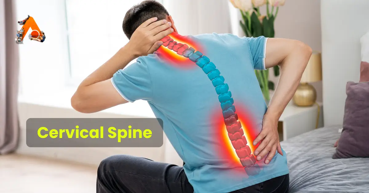
Cervical Spine
Introduction
The cervical spine plays a crucial role in supporting the head, enabling movement, and protecting the spinal cord. It consists of unique features and structures that distinguish it from other regions of the vertebral column. This article explores the anatomy, joints, ligaments, and muscles of the cervical spine while emphasizing its function and importance.
Major Functions of the Cervical Joint
The cervical spine performs the following vital functions-
- Support and Cushioning: It supports and cushions loads to the head and neck while allowing for rotation.
- Protection: It protects the spinal cord that extends from the brain.
The cervical spine is subjected to extrinsic factors such as repetitive movements, whole-body vibrations, and static loads.
Distinguishing Features of Cervical Vertebrae
The cervical vertebrae are uniquely characterized by-
- Triangular Vertebral Foramen
- Bifid Spinous Process: The spinous process splits into two distally.
- Transverse Foramina: These are holes in the transverse processes that provide passage to the vertebral artery, vein, and sympathetic nerves.
Atlas (C1)
The atlas is the first cervical vertebra and articulates with the occiput of the head and the axis (C2).
Key Features of the Atlas:
- Lateral Masses: Connected by anterior and posterior arches. Each lateral mass contains:
- Superior articular facet (for articulation with occipital condyles).
- Inferior articular facet (for articulation with C2).
- Anterior Arch: Contains a facet for articulation with the dens of the axis, secured by the transverse ligament.
- Posterior Arch: Contains a groove for the vertebral artery and C1 spinal nerve.
Axis (C2)
The axis is easily identifiable due to its dens (odontoid process), which extends superiorly from the anterior portion of the vertebra.
Key Features of the Axis:
- The dens articulates with the anterior arch of the atlas, creating the medial atlanto-axial joint.
- This joint allows for the independent rotation of the head relative to the torso.
Joints of the Cervical Spine
The cervical spine comprises two types of joints:
Joints Present Throughout the Vertebral Column
- Disc Joint:
- Located between vertebral bodies.
- Made of fibrocartilage (cartilaginous joint, symphysis).
- Functions:
- Bears the body’s weight above it.
- Provides motion, contributing to 25% of the spine’s height and 40% of the cervical spine’s height.
- Facet Joint:
- Articulation of superior and inferior articular processes from adjacent vertebrae (synovial joint).
- Also known as zygapophyseal or Z joints.
- Functions:
- Guides motion at segmental joint levels.
- The plane of the cervical facets is approximately 45 degrees, resembling a roof slope.
Ligaments of the Cervical Spine
Craniovertebral Ligaments
- Anterior Atlanto-Occipital Membrane: Connects the foramen magnum to the atlas; continues with the anterior longitudinal ligament.
- Apical Ligament: Short ligament attaching to the anterior part of the foramen magnum.
- Alar Ligaments:
- Inserted onto the occipital condyles.
- Limit axial rotation between the occiput and atlas.
- Trauma or inflammatory diseases can damage these ligaments, increasing axial rotation.
- Membrane of Tectoria: Extends from the posterior surface of the axis body to the basiocciput.
- Transverse Ligament of the Atlas: Secures the dens to the anterior arch of the atlas.
- Accessory Atlanto-Axial Ligaments
- Posterior Atlanto-Occipital Membrane
- Lateral Atlanto-Occipital Ligaments
Lower Cervical Ligaments
- Anterior Longitudinal Ligament: Lies anterior to vertebral bodies; relaxed in flexion and taut in extension.
- Posterior Longitudinal Ligament: Lies posterior to vertebral bodies in the vertebral canal; stretches in neck flexion and relaxes in extension.
- Ligamenta Flava: Connects laminae of adjacent vertebrae; allows flexion and prevents hyper-flexion.
- Ligamentum Nuchae:
- A fibroelastic membrane extending from the occiput to cervical spines.
- Provides head and neck stability, especially during flexion and acceleration injuries.
Muscles of the Cervical Spine
Posterior Muscles
- Trapezius (Traps):
- Most superficial; extends from the occiput to the lower thoracic spine.
- Functions: Neck extensor, ipsilateral lateral flexion, and contralateral rotation.
- Levator Scapulae:
- Deep to traps; extends from the first four cervical vertebrae to the scapula.
- Functions: Scapular elevation and ipsilateral lateral flexion/rotation.
- Splenius Capitis and Cervicis: Prime movers of neck and head (extension and rotation).
- Semispinalis Capitis and Cervicis: Deepest posterior muscles.
Lateral Muscles
- Scalene (Anterior, Medial, Lateral):
- Functions: Flexion, lateral flexion, and stabilization of the cervical spine.
- Important anatomical relationship: The brachial plexus, subclavian artery, and vein pass between the anterior and middle scalene muscles.
- Sternocleidomastoid (SCM):
- Extends from the manubrium and clavicle to the mastoid process.
- Functions: Lower cervical flexion, head extension, ipsilateral lateral flexion, and contralateral rotation.
Anterior Muscles
- Deep Craniocervical Flexors: Longus capitis, longus colli, rectus capitis anterior, and rectus capitis lateralis.
- Provide dynamic support to the cervical spine.
- Work with SCM for cervical flexion.
- Mandibular Elevator Group: Masseter, temporalis, and internal pterygoid muscles.
Conclusion
The cervical spine is a highly specialized structure that supports the head, enables movement, and protects the spinal cord. Its unique features, intricate joints, robust ligaments, and powerful muscles make it an essential part of the body. Understanding its anatomy and function is vital for diagnosing and treating conditions affecting the neck and upper spine.

